ABOUT FIDSSA

Case of the Month

July 2020

L. Richards MBBCh, Mmed(wits), FCP(SA)
J Nel MBChB, FCP(SA), Cert ID(SA), DTM&H
Department of Internal Medicine, division of Infectious Diseases, Helen Joseph Hospital

Photosensitive disorders in HIV

Case presentation
A 48-year-old female presented to hospital in February 2020 with a 1-week history of ‘generalised body weakness’ and diarrhoea. No haematochezia was noted. In addition, she had been coughing for 3 weeks and this was associated with loss of weight and night sweats. On further enquiry she complained of a 1- year history of a rash on her face, neck and arms. It started as raised, dark areas and progressed to its current state. It had never been pruritic or painful. She had no history of joint pain or stiffness, no Raynaud’s phenomenon, no mucosal ulceration, no sicca symptoms or alopecia. She had no animal exposure or travel history. She had never used any skin-lightening products and had no known allergies.

The patient was HIV reactive and is currently on zidovudine 300mg daily, lamivudine 300mg daily and lopinavir/ritonavir 400mg/100mg 12-hourly which was started on January 3rd 2020. Her antiretroviral treatment (ART) history prior to this is unclear, although it was thought that she was on tenofovir DF/emtricitabine/efavirenz fixed dose combination tablet previously. The patient was admitted to another hospital in December 2019 with a right cerebrovascular accident (CVA). Her other chronic medication includes aspirin and simvastatin started after her CVA.

On admission her vital signs were: a heart rate of 128b/min, blood pressure 92/63mmhg, she was pyrexial with a temperature of 38.2°C, her respiratory rate was 20b/min and a random glucose was 8.2mmol/L. Generally, she was wasted and chronically ill looking. She had hypopigmented, vitiligo-like, erythematous depigmentation of her face, neck and arms. (Figures 1-4) There was some skin scaling around these areas. She had no significant lymphadenopathy. On auscultation of her chest she had decreased breath sounds in the left lower zone. She has a residual left hemiplegia on her neurological exam with a power of 3/5, increased tone and hyperreflexia. Her cardiovascular and abdominal examination were unremarkable.

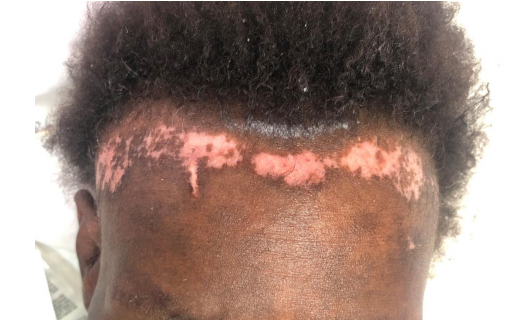
Figure 1: The patient’s rash on her forehead

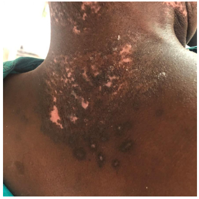
Figure 2: The patient’s rash on posterior neck and upper back

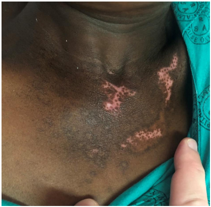
Figure 3: The patient’s rash on her chest

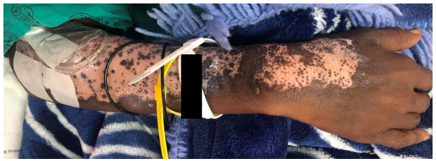
Figure 4: The patient’s rash on her right forearm

She had a CD4 count of 5 cells/µL, an HIV viral load of 287 000 copies/mL and a negative serum cryptococcal antigen. Her sputum GeneXpert MTB/Rif Ultra came back positive for M. tuberculosis, with rifampicin sensitivity

What is the differential diagnosis of this patient’s skin rash?

Photosensitive drug reactions can be ‘phototoxic’ or ‘photoallergic’. Phototoxic reactions look like a severe, well-demarcated sunburn, occurs within minutes to hours of sun exposure and causes a burning sensation. Photoallergic reactions are a type IV hypersensitivity reaction occurring 24-48 hours after sun exposure. It is usually eczematous in appearance and is associated with pruritus. Some of the drugs implicated in this syndrome are sulphonamides, NSAIDs, pyridoxine and efavirenz1. This could be a possibility in our patient as she may previously have been on efavirenz or cotrimoxazole. Factors against this diagnosis are the fact that it did not start out as a well demarcated rash, it is not eczematous, was not pruritic or painful and it has not improved since stopping these medications (if she were ever on them).

Chronic actinic dermatitis (CAD) includes a spectrum of conditions including photosensitive eczema, actinic reticuloid photosensitivity eczema and persistent light reaction. These were previously thought to be separate entities but are now considered a continuum of CAD. It is usually present in men over the age of 50 years. It presents as thickened, scaling patches and plaques which become confluent and can involve sun unexposed areas. It can heal with depigmentation 1;2. This diagnosis could apply to our patient in that she does describe the rash starting as dark, raised areas and it has progressed to a depigmented lesion. It is more common in older men however and has not progressed to involve sun unexposed areas.

Actinic lichenoid leukomelanoderma of HIV is a condition that has only ever been described in South Africa in patients with low CD4 counts. It presents as symmetrical, well circumscribed, scaly plaques which are hyperpigmented and violaceous or blue-grey in colour. It progresses to a centrally depigmented or erythematous center with hyperpigmented peripheries. It is initially pruritic and can resemble discoid lupus erythematosus 1;3. This diagnosis fits nicely with our patient aside from the symptom of pruritus.

Discoid lupus erythematosus mostly involves the face, neck, scalp and ears. It less frequently involves the torso. The lesions present as erythematous papules or plaques covered by a scale. As it progresses, the scale will thicken and become adherent. Lesions can become hypopigmented centrally with hyperpigmented edges and heal by leaving an atrophic central scar 4. The antinuclear antibodies (ANA) and extractable nuclear antigen (ENA) results were negative in our patient and she had no other features to suggest SLE. She did not have any atrophic scarring.

Lichenoid photoeruptions are violaceous to slatey grey, flat-topped papules and plaques which occur on the face, dorsal forearms, hands and lower lip. It resembles lichen planus but spares the mucosa 1. This may have fitted our patient in the early stages of the rash but now that she has hypopigmentation it does not fit the clinical picture of a lichenoid photoeruption.

Management

The patient was started on rifampicin, isoniazid, pyrazinamide, ethambutol and pyridoxine for her pulmonary tuberculosis. Her lopinavir/ritonavir dose was doubled and a skin biopsy was performed. She was asked to follow up with dermatology in 4 weeks-time.

Final diagnosis

The skin biopsy confirmed a diagnosis of actinic lichenoid leukomelanoderma of HIV.

Discussion

It is estimated that 5% of HIV reactive patients have some form of photosensitive dermatitis. The differential diagnoses include photosensitive drug reactions, CAD, pellagra, lichenoid photoeruptions, porphyria cutanea tarda, pseudoporphyria, photoaggravated granuloma annulare, actinic prurigo and actinic lichenoid leukomelanoderma1.

The term actinic lichenoid leukomelanoderma (ALLMD) was first used by Wolfowitz and colleagues at the South African Dermatological Society’s congress in 2005. They described a photosensitive skin reaction associated with advanced HIV infection, lichenoid features with pigment incontinence on histology and not associated with medication. Unfortunately, their data was not published3. To our knowledge this condition has only been reported in South Africa and only one publication describes it1. In an unpublished case series of 12 patients diagnosed with ALLMD they all had low CD4 counts, the highest being 53 cells/µL. The authors of this case series offer a therapeutic regimen that seemed to improve the symptoms of those patients that followed up. This includes3:
  • Antiretroviral therapy
  • Chloroquine 200mg daily
  • Intermediate potency corticosteroids for facial lesions and ultra-potent corticosteroids for lesions on the trunk given until the lesions start to flatten out
  • Sun protection
HIV reactive patients are more susceptible to photosensitive disorders often healing with disfiguring scars. In order to manage these patients to prevent permanent damage they should be investigated to establish a diagnosis. Involve a dermatologist early on, consider medications as a cause and investigate for a systemic disorder.

Recommended reading

  • Koch K. Photosensitive disorders in HIV. S Afr J HIV Med. 2017;18(1): 676 doi: 10.4102/sajhivmed.v18i1.676
  • Gregory N, DeLeo VA. Clinical manifestations of photosensitivity in patients with human
    immunodeficiency virus infection. Arch Dermatol. 1994;130(5):630-633.
    doi:10.1001/archderm.1994.01690050098018
  • Vin-Christian K, Epstein JH, Maurer TA, McCalmont TH, Berger TG. Photosensitivity in HIV-infected individuals. J Dermatol. 2000;27(6):361–369.
    doi.org/10.1111/j.1346-8138.2000.tb02185.x

Earn CPD Points

CPD QUESTIONS

FIDSSA Members can earn CPD points by logging into the secure section of the website and visiting the MyCPD section.