ABOUT FIDSSA

Case of the Month

October 2019

Dr Max Peter Winkler
South African Society of Travel Medicine

Case Report 1

A 48-year-old male patient presented in July 2019 with a one-week history of an erythematous rash on the right thigh, arthralgia, myalgia and persistent headaches. The symptoms had started soon after he had returned with his family from a holiday to the Scottish Highlands (Kyle District). He had spent a large amount of time during the holiday hiking with his family in the Scottish countryside and noted that he had been bitten by a tick while out hiking. He is originally from the United Kingdom but now lives in Cape Town with his family. He travels extensively for work and has no other medical co-morbidities and is not currently on any chronic medication.

On examination a large flat (+- 5cm) erythematous skin lesion was noted on the lateral aspect of the right upper thigh. There was no evidence of an eschar or lymphadenopathy in the vicinity of the bite. He was apyrexial at the time of examination and he had no other physical findings of clinical significance.

Case Report 2

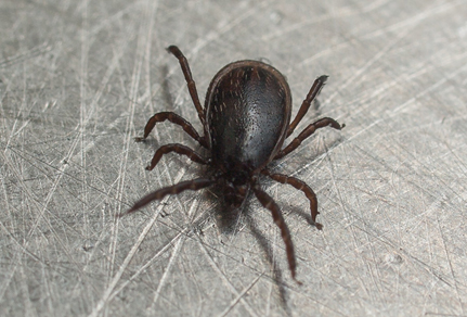
A 47-year-old male patient presented in July 2019 with a one-week history of arthralgia, myalgia, headache and a slight fever. He had recently travelled to Los Angeles (USA) for a work conference where he was bitten by an unknown insect in the right axilla while staying in rented accommodation. He mentioned that he did find some ticks in the garden of the house at the time of his stay. He stopped off in the UK and Holland on his way back to South Africa, but he did not spend any time outdoors while there. He is originally from Zimbabwe but has lived in South Africa for the past 20 years. He has no co-morbid illnesses and is not currently on any chronic medication.

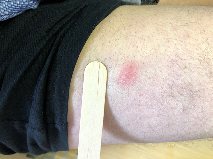
On examination a small punctate skin lesion was noted in the right axilla. However, there was no surrounding erythema or eschar noted at the bite site and no associated lymphadenopathy. He was apyrexial at the time and the rest of his physical examination was normal.

Differential Diagnosis

With the history of tick exposure and the findings on physical examination presented above we should consider the following in our differential diagnosis for these two cases.

  • Rickettsia: these infections are caused by bacteria from the order Rickettsiales and are classically divided into the spotted fever and typhus groups (1). The spotted fevers are typically spread by tick bites and are common in travelers returning from outdoor holidays and safaris. However, the rash in Case 1 is not typical of these infections.

  • Tickborne Encephalitis (TBE): this disease is caused by a flavivirus and is transmitted to humans through the bite of infected ticks from the Ixodes species in Europe and the far east (1). Even though the clinical presentation in these cases might be similar to TBE there have been no reported cases of endemic TBE in the UK or the USA, and the patients gave no history of recent travel to central Europe or Asia therefore this diagnosis is unlikely.

Final Diagnosis

Introduction
Lyme disease was first described in 1975 during an outbreak of cases in the town of Lyme, Connecticut from where the name is originates. It is caused by spirochetes from the Borrelia family specifically the Burgdorferi families in North America and the Afzelli and Garinii families in Europe and Asia.

Distribution

The most common vectors for borrelia transmission in North America and Europe are the Ixodes (blacklegged) ticks. These ticks are distributed across Europe from Scandinavia in the north and the Mediterranean in the south to the British Isles in the west and central Russia in the east. Endemic areas in North America are the north eastern and north central United States and Canada.

In recent years distribution has spread across North America and ticks have been found outside their historical range in previously non-endemic areas such as California. In Asia, infected Ixodes ticks are found from Russia through Mongolia to northeastern China and Japan although human infection is uncommon in Asia (1).

Figure 2: Global distribution of Ixodes ticks (2)

The nymphal stage of the tick is responsible for most bites in humans. These nymphs are approximately the size of a poppy seed and are very difficult to detect. They mostly feed on small mammals such as mice and other rodents and are most active from May-July when the temperatures start to warm in the northern hemisphere (2).

Figure 3: Developmental stages of the Ixodes tick (3)

Signs

The rash described in Case 1 is an example of erythema migrans on the lateral aspect of the right thigh. Erythema migrans is diagnostic for Lyme disease: the rash may be flat or slightly raised and often appears after 1-33 days (on average 7-10 days) at the sight of a tick bite, and typically fades after 3-4 weeks if not treated. The rash is generally circular but may be linear or triangular and often a central punctum is present at the bite site. The rash enlarges by a few centimeters per day and most lesions will achieve an average diameter of +- 15cm, but some as large as 70 cm have been described in the literature. The entire lesion may be uniform in colour or show central darkening, though central clearing may also develop (more commonly in European than North American cases). There may be additional erythema proximal to the lesion giving rise to the so-called bull’s eye appearance, but this only occurs in a minority of patients (4).

Figure 1: Rash on the right lateral thigh of Case 1.

Symptoms

Complications in multiple organ systems have been described in Lyme disease. The incubation period ranges from 3-30 days after exposure to an infected tick bite with 80% of patients developing acute erythema migrans at the bite site. This is pathognomonic of Lyme infection Other early symptoms may include the following: fatigue, headache, myalgia and lymphadenopathy. If left untreated further symptoms may develop in the following weeks to months. These symptoms may involve multiple organ systems and can often be difficult to distinguish from other more common ailments which may pose a diagnostic dilemma for the clinician, especially if there is no definitive history of recent tick exposure. The longer-term symptoms may include the following:

  • Neurological complications including meningitis, radiculopathies, facial palsies, neuropathies and encephalopathies.
  • Cardiac abnormalities including myocarditis with AV node block.
  • Chronic monoarticular or oligoarticular arthritis affecting multiple joints.
  • Chronic skin rashes such as acrodermatitis chronica atrophicans and lymphocytoma (these seem to be more common in European borrelia infections)

Figure 4: Acrodermatitis chronica atrophicans (web reference:  Click here).

Investigations

Serology followed by confirmatory western blot testing is recommended in suspected cases. However, if the erythema migrans rash is present then the need for further testing before commencing antibiotic treatment might not be necessary.

If the patient has other symptoms suggestive of Lyme disease and a history of tick exposure, one can proceed to laboratory testing to confirm infection. This should start with IgM and IgG serology. If the serology is positive then exposure should be confirmed with a western blot panel to identify borrelia infection. If both tests are negative, and the patient is still symptomatic after 4-6 weeks, repeat testing should be considered.

The final serology and western blot results for the case studies are shown in the table below. Rickettsia serology results were included for comparison.

  • Case 1: IgM serology and western blot were both positive for Lyme disease. This result confirms a case of acute Lyme disease in this patient. The diagnosis corresponds well with the clinical presentation described, especially the presence of erythema migrans on the right thigh.
  • Case 2: IgM serology was positive, but the western blot result was reported as indeterminate Therefore acute Lyme disease cannot be confirmed in this patient. However, the diagnosis is still a possibility and should be followed up with repeat testing in 4-6 weeks to see if the western blot result changes.

Treatment

Doxycycline as well as amoxicillin and azithromycin are all indicated as oral treatments for uncomplicated Lyme disease. The majority of patients with uncomplicated disease (erythema migrans) can be treated with oral antibiotics alone. High doses of parenteral ceftriaxone should be considered for complications involving the CNS, CVS or skin. Note that higher than standard doses of ceftriaxone and doxycycline are required for CNS involvement. Treatment duration ranges from 21-28 days depending on the presence of complications (5).

Figure 5: NICE guidelines for the treatment of Lyme disease (5).

Conclusion

Both cases were started on doxycycline 100mg BD for 21 days and both have recovered fully and not reported any chronic symptoms after completion of their treatment.

Even though Lyme disease is not considered to be endemic in South Africa, one should be aware of it as a possible diagnosis in travellers from the northern hemisphere who present with typical symptoms such as erythema migrans and a history of possible tick exposure while travelling.

References

  1. Brunette GW, Nemhauser JB. CDC Yellow Book 2020. Oxford University Press; 2019.
  2. Stanek G, Wormser GP, Gray J, Strle F. Lyme borreliosis. Lancet (London, England). 2012 Feb 4;379(9814):461–73.
  3. Schotthoefer AM, Frost HM. Ecology and Epidemiology of Lyme Borreliosis. Clin Lab Med. 2015;35(4):723–43.
  4. Dainsberg C. Recognizing lyme disease. Am J Nurs. 2018;118(7):13.
  5. Cruickshank M, O’Flynn N, Faust SN. Lyme disease: Summary of NICE guidance. BMJ. 2018;361(April):1–6.

Earn CPD Points

CPD QUESTIONS

FIDSSA Members can earn CPD points by logging into the secure section of the website and visiting the MyCPD section.