
Case courtesy of: Nithendra Manickchund, Pariva J. Chutterpaul, Sonal R. Verma, Bernadett I. Gosnell, M. Yunus S. Moosa.
Department of Infectious Diseases, Nelson R. Mandela School of Medicine and King Edward VIII Hospital, Durban.
Written consent was obtained from the patient to publish images.
A 41-year-old woman presented to King Edward VIII hospital acute medical unit with a four-month history of progressive skin lesions. The lesions started on her face and gradually progressed to involve her trunk and arms. In the three weeks prior to admission, she developed constitutional symptoms and a productive cough. An HIV ELISA test was positive; she had never tested for HIV in the past. She denied a headache, visual disturbance or photophobia.
Social History:
She had worked in Hong Kong as an entertainer at an amusement park. She had travelled at least twice a year between Hong Kong and South Africa over the past three years. The skin rash began during her last trip to Hong Kong and worsened on her return to South Africa four months ago.
She had no significant past medical history or allergies. She was not using any medication.
Examination findings:
Notable findings were a temperature 37.2oC and significant conjunctival pallor. There was discrete rubbery cervical and supraclavicular lymphadenopathy, the largest nodes measuring 2.5 cm x 2 cm.
On skin examination there were multiple ulcerating papules that coalesced to form plaques with areas of haemorrhage and scabbing. Lesions were of varying sizes and ages. Early-stage papular lesions were centrally umbilicated. Lesions involved her face, upper arms, back and chest and a few lesions were noted on both thighs.
She was not distressed, with a respiratory rate 18 breaths/minute, and crepitations in the right middle zone anteriorly.
Her liver was soft and could be palpated 3 – 4 cm below the costal margin, with a total span of 16 cm. There was no splenomegaly or ascites.
Cardiovascular and neurological examinations were normal.
Unfortunately all of the patients X-rays were lost due to problems with the porters at the hospital.
Differential: Neutrophils: 4.85 x 10⁹ (88%) Lymphocytes 0.6 x 10⁹ (10%)
Smear: Scanty teardrops, moderate anisocytosis, moderate polychromasia, dimorphic red cell picture. No yeasts seen.
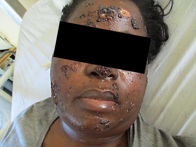
Figure 1: Lesions on the face on presentation (12 May 2018)
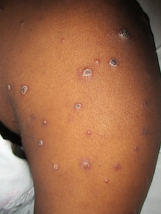
Figure 2: Lesions on the upper arm at presentation demonstrating a polymorphic rash with lesions at varying stages of evolution. (12 May 2018)
What is the differential diagnosis for this presentation in a person with advanced HIV disease with a constitutional symptoms, skin lesions and a lower respiratory tract process?
How would you investigate this patient further?
In this patient:
What is talaromycosis?
Talaromycosis (formerly penicilliosis) is an opportunistic infection caused by Talaromyces marneffei (previously known as Penicillium marneffei). This infection is caused by a thermally dimorphic fungus and the only one in this genus to cause disease in humans. It is endemic and limited to South East Asia where it is the fourth most common opportunistic infection in patients with AIDS.
When should you suspect T. marneffei infection?
The clinical features are non-specific. One needs to rely on the history and investigations to make a diagnosis.
One should suspect this infection in immunocompromised patients who give a history of travel to endemic regions in South East Asia such as Thailand, Vietnam, Hong Kong, Southern China, Taiwan, India, and Laos. Reports of patients developing the disease more than ten years after travel to South East Asia suggests that a history of both recent and remote travel is of clinical significance.
What is the natural reservoir and mode of transmission of infection?
Much remains unknown about the natural reservoir and route of transmission of T. marneffei. The only known reservoirs of the fungus are humans, bamboo rats and soil in endemic regions. Case-control studies suggest that exposure or consumption of the bamboo rat is not a risk factor. However, several reports suggest that a recent history of exposure to soil, particularly in the rainy season, is a risk factor. It has been posited that an airborne route of acquisition, through inhalation of conidia from an environmental source, followed by dissemination to other body sites during immunosuppression is the most likely pathogenesis of infection. T. marneffei infection has been described as a source of laboratory-associated infection and biosafety level 2 facilities are recommended when manipulating cultures.
How does a patient with T. marneffei present?
The signs and symptoms of disease are non-specific. At presentation, patients have generally been ill for approximately four weeks with weight loss, low-grade fever and constitutional symptoms. Skin lesions, in the form of papules, pustules, nodules, ulcers or abscesses occurring on the face, trunk and extremities, have been reported in 50%-70% of patients. In the HIV-infected person, papules with central necrosis or umbilication resembling molluscum contagiosum are commonly seen.
Other commonly seen clinical features include anaemia, generalised lymphadenopathy, hepatomegaly and splenomegaly. Involvement of the pharynx and palate has also been described, particularly in patients with AIDS. Pulmonary involvement can appear radiologically as reticulonodular, nodular, or diffuse alveolar infiltrates. Cavitation with haemoptysis has been uncommonly described. Central nervous system involvement is rare; however, at autopsy, meningeal involvement has been described. Lumbar puncture is not routinely recommended in patients who are not symptomatic.
How would you confirm a diagnosis of talaromycosis?
Culture is the gold standard for diagnosis with an almost 100% yield from bone marrow and lymph nodes. Yield from skin biopsy is approximately 90% and blood culture about 76%. (1)
Serum galactomannan antigen can be positive in 70- 80% of patients with disseminated talaromycosis as there is cross reactivity in the assays designed to detect the galactomannan of Aspergillus species with the galactomannan of Talaromyces species. However, lack of specificity limits its diagnostic value.
Microscopy is of value. Intracellular yeast cells (with central septa) can be seen within monocytes on peripheral blood smear, in fine-needle aspirates of lymph nodes, sputum cytology and touch smears of skin.
On histology, T. marneffei can be mistaken for H. capsulatum as the yeast forms are similar in size, particularly in the extracellular form. If the histopathologists were aware of the travel history in our patient, they would have considered T. marneffei in their differential diagnosis. However the travel history was not evident until later in her hospital stay.
T. marneffei divides by central septate fission rather than budding. Therefore the absence of budding and the presence of a central transverse septum can be used to distinguish it from H. capsulatum on histology. It also tends to be more elongated than H. capsulatum.
Diagnosis may be challenging in an immunocompetent patient as fungal staining may be negative and granulomatous inflammation may be the only histological finding. Culture is required for full identification although molecular methods are newly available.
The culture medium of choice is Sabouraud agar where growth in the form of mycelia is seen within two to five days at 30 degrees Celsius. Colonies are grey and typically produce a characteristic red pigment which diffuses into the agar.
Unexpectedly, despite T. marneffei existing in yeast form at 37°C, it can appear as septate hyphae-like structures on Gram stain of positive blood cultures, particularly if the blood culture bottle is kept at room temperature before the Gram stain is prepared.
In our patient, we became suspicious of a diagnosis of talaromycosis when the patient’s travel history was obtained by the general medical team. A fungal aetiology became more apparent when we were alerted by the microbiologist that hyphae-like structures were noted on the Gram stain of the blood culture that flagged positive. However, the production of diffusing red pigment on culture of a skin biopsy taken by the dermatologist on admission, as well as the blood culture virtually clinched the diagnosis prior to full identification and molecular confirmation.
How would you manage this patient?
The mortality from disseminated T. marneffei is 100% if untreated. A delay in the initiation of therapy is associated with poorer outcomes.
In a case series of HIV-infected patients, amphotericin B (AMB) and itraconazole were shown to be equally effective with a response rate of 77% and 75% respectively. (1) Currently it is recommended that intensive treatment with AMB (AMB deoxycholate 0.6 mg/kg or liposomal AMB 3-5 mg/kg) should be given for 2 weeks followed by oral itraconazole 400 mg/day for 10 weeks (2).
Milder forms of the disease can treated just with oral itraconazole 400 mg/day for 8 weeks (2). Fluconazole has poor activity with clinical response rates as low as 30% (1). Voriconazole is considered an acceptable alternative (2).
Prior to the availability of antiretroviral therapy (ART), the relapse rate was over 50% in the first six months following completion of treatment. Itraconazole secondary prophylaxis (200 mg/day) reduced the relapse rate from 57% to 0%. Current guidelines recommend secondary prophylaxis until the CD4 cell count increases above 100 cells/ mm3 for at least six months. (2,3)
There are no studies addressing the timing of initiating ART. Guidelines have used information from clinical trials and anecdotal experience with other opportunistic infections. Currently it is recommended to start ART as soon as possible after starting anti-fungal therapy in patients with a CD4 count of less than 50 cells/mm3. ART can be delayed until completion of the two weeks’ induction therapy in patients with a CD4 count more than 50 cells/mm3. (2)
Our patient developed shortness of breath and became distressed within three hours of the infusion of the first dose of AMB deoxycholate. Pulmonary reactions are a rare but recognized adverse effect of AMB, likely related to the rapid infusion (4). Our patient had received the infusion rapidly as it was almost complete at the time of developing the symptoms and likely contributed to the severity of the reaction. She became hypoxic and confused with bilateral diffuse crepitations and wheezes on auscultation of her chest. Arterial blood gas testing revealed a type II respiratory failure and chest X-ray showed new bilateral diffuse infiltrates. The intensive care unit deemed her unsuitable for management in the ICU. AMB was discontinued and she was commenced on intravenous hydrocortisone 100 mg 8 hourly. Within 48 hours, she made a remarkable recovery from her respiratory symptoms.
It was decided not re-challenge with AMB and instead, intravenous voriconazole was commenced. She was subsequently transitioned to oral itraconazole, which broadly covered for all fungal possibilities on the differential diagnosis.
What are the concerns with use of itraconazole in the South African context?
Itraconazole as an oral agent is available in a solution and capsule form. The itraconazole solution preparation has a more reliable absorption with an enhanced bioavailability and has to be taken on an empty stomach. Absorption of the capsule preparation is dependent on a low gastric pH and is enhanced by food or cola beverage. Itraconazole is only available in South Africa in an oral/capsule formulation.
The major concern with itraconazole therapy in the current SA context is drug-drug interactions with both the current first and second line ART regimens. The non-nucleoside reverse transcriptase inhibitors (NNRTIs) cause a significant reduction in itraconazole levels. The protease inhibitors (PIs) cause a significant increase in the itraconazole levels due to inhibitory effects on the cytochrome P450 enzymes. Kristel et al 2004 showed that even after halving the recommended dose of itraconazole from 400 mg to 200 mg daily, supra-therapeutic levels of the drug were recorded due to reduced metabolic degradation secondary to CYP3A4 inhibition by lopinavir/ritonavir.
In the absence of therapeutic drug monitoring, dosing of itraconazole is challenging. We decided to use the pharmacokinetic effect of the protease inhibitors to ensure that we achieve adequate serum levels of itraconazole and monitored her closely for adverse events such as abnormal liver enzymes, lipid abnormalities, electrolyte abnormalities and haematological abnormalities.
We started itraconazole 200 mg three times daily for 3 days followed by twice daily for 2 days. We reduced the dose to 200mg daily on starting ARV’s. This was 1 week into treatment and she was initiated on tenofovir/emtricitabine/lopinavir/ritonavir
Our patient responded very well with no adverse drug reactions. Her CD4 cell count increased to 258 cells/mm3 and HIV-1 viral load dropped to 164 RNA copies/ mL (2.21 log) after ten weeks of ART. Her cutaneous lesions showed remarkable improvement (Figure 3).
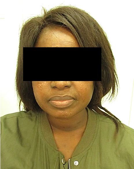
Figure 3: Improvement in skin lesions after 10 weeks on itraconazole treatment (200 mg daily)
(6 August 2018)
Acknowledgements
We acknowledge the contribution made by Dr. P. Mahabeer from the Department of Microbiology NHLS. We also acknowledge the contribution made by Dr Ameshin Moodley, Dr Nerissa Moodley of the Department of Dermatology, King Edward VIII Hospital, Durban for assisting in the diagnosis.
References:
FIDSSA Members can earn CPD points by logging into the secure section of the website and visiting the MyCPD section.
Atlasville, Boksburg
South Africa
2022 © FIDSSA - All rights reserved • Website Terms of Use • Privacy Policy • Powered by E2
Admin login | Website login |
MYMEMBERSHIP®