ABOUT FIDSSA

Case of the Month

December 2019

Unexplained hepatitis in a returning traveller, a case report on Hepatitis E

Dr A Engelbrecht MB ChB, Dip HIV Man (SA)
Dr MY Brinkhuis MB ChB
Dr JL Pretorius MB ChB, Mmed (internal Medicine and Infectious Diseases), FCP SA
Department of Internal Medicine, Worcester Hospital

Case Presentation
Our patient was a 61-year-old female from South Korea. She presented with a hepatitis, giving a 4-day history of fever, rigors, nausea, vomiting, myalgia and headache. She recently travelled to the Philippines, South Korea and Kenya before arriving in South Africa. She attended a work-related conference in Manilla in the Philippines 3 weeks prior to presentation, during which time she stayed in a conference hotel. She denied any sexual activity, alcohol use, tobacco, or illicit drugs. She denied taking any over the counter or prescribed medicines or herbal supplements. She only ate the hotel food and drank bottled water. She returned to South Korea for 3 days before leaving for Kenya. She arrived in Nairobi 1 week prior to presentation. She only stayed in the capital city with no travel to rural areas. Again, only eating hotel food with no specific insect or rodent exposure. She received no previous blood transfusions and had no personal or family history of liver disease.

Admission vital signs were in the normal range. On examination she was apyrexial, not jaundiced, had no eschars or rashes, and had mild epigastric tenderness with no organomegaly and no clinical features of liver failure. After 2 days in a general medical ward she became icteric. Her blood results are summarized in Table 1.

Table 1
(Abbreviations for Table 1 - TB: Total Bilirubin, CB: Conjugated Bilirubin, ALT: Alanine aminotransferase, AST: Aspartate aminotransferase, ALP: Alkaline phosphatase, GGT: Gamma-glutamyl transpeptidase, LDH: Lactate dehydrogenase, INR: International Normalised Ratio)

Further testing revealed negative HIV ELISA, negative Hepatitis A IgM, negative Hepatitis B surface antigen (HepBsAg) and surface antibody (HepBsAb), negative Hepatitis C IgG, and negative malaria thin and thick smear. An ultrasound of the liver was performed and showed normal liver structure and vasculature with a normal biliary system and no lymphadenopathy.

What is the Differential Diagnosis? Non-Infectious:
  • Auto-immune Hepatitis
  • Drugs
Infectious:
  • Viral Hepatitis (A, B, C, E)
  • Yellow Fever
  • Leptospirosis
  • Typhoid

Management Antinuclear antibody (ANA) was negative. Patient denied any drug use.

Viral Hepatitis A was excluded with negative IgM. Hepatitis B surface antigen (HepBsAg) and surface antibody (HepBsAb) were negative. However, HepBsAb may in certain cases not be detectable until after a window period of several weeks to months, during which time neither HepBsAg nor HepBsAb can be detected (figure 1)[1]. At this time, the serologic diagnosis can be made by the detection of IgM antibodies against hepatitis B core antigen (anti-HepBc IgM) [1]. Our patient subsequently had a negative anti-HepBc IgM.

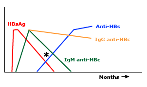
Figure 1: Window period of acute HBV infection [1]
(© 2019 UpToDate, Inc. and/or its affiliates. All Rights Reserved)

The National Institute for Communicable Diseases (NICD) was consulted regarded the possible diagnosis of yellow fever. Yellow fever is a vector-borne acute viral haemorrhagic disease caused by an infection with the Yellow Fever virus (YFV). The virus is transmitted by a bite of an infected mosquito vector Aedes aegypti [2]. The laboratory diagnosis of yellow fever is based on the detection of YFV antigen through PCR and serologic assays [3]. The NICD reviewed the patient’s travel history and safely excluded the diagnosis based on no yellow fever risk in the areas she visited. Although yellow fever vaccination is recommended for Kenya, there is specifically no risk in Nairobi (figure 2).

Figure 2: Yellow fever vaccine recommendations in Africa [3]

Leptospirosis serology was negative.

The patient was empirically treated with Ceftriaxone for 5 days for suspected typhoid fever. Blood cultures failed to isolate any pathogens. Although previous literature review on the sensitivity of blood culture for Salmonella Typhi suggested only 40 to 60 % [4], we felt the negative blood cultures together with the clinical constellation of no fever, no abdominal pain and no diarrhoea, made the diagnosis of typhoid fever unlikely.

What is the final diagnosis?

Acute hepatitis E with hepatitis E PCR and IgM positive. Genotyping revealed that the infected virus belongs to the HEV genotype 4 based on phylogenetic analysis. It seems to suggest that the patient acquired the hepatitis E either in South Korea or in the Philippines, and not in Kenya (see figure 3).

Discussion

Hepatitis E was first suspected 30 years ago in Kashmir India where a waterborne outbreak of hepatitis occurred and was found not to be related to Hepatitis A or Hepatitis B virus. At the same time a similar outbreak happened in New Delhi where serology also did not demonstrate either Hepatitis A or Hepatitis B. The confirmation of these observations came in 1983 through demonstration at immune electron microscopy of spherical virus-like particles in stool specimens collected from a human volunteer who had ingested a pooled faecal suspension from patients with acute hepatitis in Afghanistan. [5]

Hepatitis E has a global distribution. It falls in the genus Hepevirus and family Hepeviridae. Based on studies performed on viral genomic sequence, at least 4 genotypes have been described, with one shared serotype. There are two distinct epidemiological patterns. In developing countries such as Asia and Africa with high disease prevalence, it can occur as outbreaks or sporadic cases of acute hepatitis exclusively related to Genotype 1 and 2. The route of transmission is usually faecal-oral in areas with poor sanitation and subsequent water contamination. In developed countries with lower disease prevalence and better water and sanitation supply, sporadic cases of locally acquired Genotype 3 and 4 have been reported. The route of transmission is often zoonotic transmission to humans from consumption of undercooked meat, specifically pork. [5]

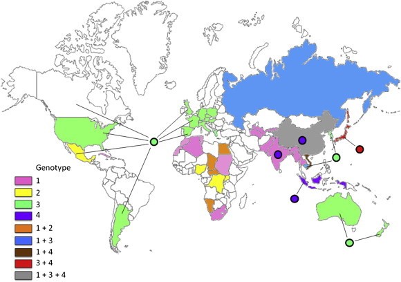
Figure 3: Distribution of HEV genotypes in viral isolates obtained from humans and animals (mainly pigs). The colors used for a country and the circle associated with it represent the predominant HEV genotypes of human and animal isolates, respectively, from that country. The figure is based on data from Okamoto, 2007 [5]

The World Health Organization (WHO) estimate that 20 million HEV infections occur annually, with over 3 million symptomatic cases and in excess of 55 000 HEV‐related deaths, accounting for 3.3% of the total mortality due to viral hepatitis [6]. According to the Centers for Disease Control and Prevention (CDC) mortality in the third trimester of pregnancy reaches 10%–30% [7]. They have found that equally high mortality rate occurs in solid organ transplant recipients on immunosuppressive therapy and in patients with pre-existing chronic liver disease [7].

Recent prospective studies in 2016-2017 conducted by the University of Cape Town and Stellenbosch University respectively, concluded that the HEV IgG seroprevalence in the Western Cape, South Africa is 27.9% and 26% [8]. This indicates that the prevalence of HEV as a cause of acute hepatitis in the Western Cape is higher than previously thought.

HEV has an incubation period of 3 to 8 weeks, followed by a short prodromal phase followed by jaundice lasting days to several weeks [9]. Acute HEV can be diagnosed with the detection of anti-HEV IgM, which is often detectible on day 4 after the onset of jaundice and can persist for 3–5 months. The gold standard for the diagnosis of active HEV infection is detection of HEV RNA [10].

HEV infection is generally self-limiting in immunocompetent patients, as was the case in our patient. However hepatitis E can cause chronic hepatitis in immunocompromised patients and needs to be treated. Treatment mainly consists of reducing the dosage of immunosuppressive therapy to help in the clearance of HEV. Ribavirin has also been shown to be effective in solid organ transplant recipients and in patients with persistent chronic infections [11].

With a seroprevalence of almost 28% in the Western Cape, we would like to propose that hepatitis E should not only be considered in any traveller presenting with hepatitis, but also in local patients presenting with an unexplained hepatitis.

Recommended Reading

  • RG Madden, S Wallace, M Sonderup, S Korsman, T Chivese, B Gavin, A Edem, R Govender,N English, C Kaiyamo, O Lutchman, et al. Seroprevalence of hepatitis E virus (HEV) in Western Cape, South Africa. Journal of Viral Hepatitis 2015 John Wiley & Sons Ltd, Vol 22 (Suppl. S2), June 2015, 19–135.
  • Korsman S, Bloemberg J, Brombacher M, Giuricich A, Halley-Stott RP, Kaba M. Hepatitis E In pig-derived food products in Cape Town, South Africa, 2014. South African Medical Journal. 2019 Jul 26;109(8):584-586. doi: 10.7196/SAMJ.2019.v109i8.13868.
  • Korsman S, Hardie D, Kaba M. Hepatitis E virus in patients with acute hepatitis in Cape Town, South Africa, 2011. S Afr Med J. 2019 Jul 26;109(8):582-583. doi: 10.7196/SAMJ.2019.v109i8.13867.
  • Mohammad S Khuroo, Mehnaaz S Khuroo, Naira S Khuroo. Hepatitis E: Discovery, global impact, control and cure. World J Gastroenterol 2016 August 21; 22(31): 7030-7045
  • Ankcorn MJ & Tedder RS. Hepatitis E: the current state of play. Transfusion Medicine, 2017, 27, 84–95. Pubmed.
  • World Health Organization. Hepatitis E Fact sheet (updated July 2016). http://www.who.int/mediacentre/factsheets/fs280/en/  (Accessed on June 20, 2017)

Earn CPD Points

CPD QUESTIONS

FIDSSA Members can earn CPD points by logging into the secure section of the website and visiting the MyCPD section.