
Case presentation
Max Winkler, SASTM
In March 2018 a patient presented to a health care facility in Hout Bay, Cape Town after having been bitten by a large male brown fur seal at the local harbour earlier that day. He had travelled with his wife from the northern suburbs of Cape Town to spend the afternoon sightseeing and eating fish and chips at the picturesque local fishing harbour. After lunch they decided to take a stroll along the quay and while walking along the quayside he noticed a local resident feeding fish to a large male fur seal. This practice is common amongst residents, who entice visitors to feed the seals and then charge the tourists a small fee. According to the patient, the fur seal reared up and bit him on the inner aspect of his right thigh as he was walking past the animal.
What is the distribution of the fur seal in South Africa?
Seals, including fur seals, are members of the pinniped family with the most notable distinguishing feature being the presence of external ears in fur seals and internal hidden ears in true seals. In South Africa, brown fur seals are the predominant species of pinniped found along our coastline and their habitat extends from Cape Cross in Namibia to Port Elizabeth in the Eastern Cape where they breed on rocky outcrops and small offshore island. Near the base of sentinel peak in Hout bay on a small rocky island off the coast (Seal Island) there is a large breeding colony of brown fur seals. These seals often visit the local harbour to forage for fish and sun themselves on the quay. The majority of bites (n=8) have occurred in the harbour on the quayside involving a tourist and local seal “tamer” trying to entice visitors to take photos and feed the seals for a small fee. However there have also been some incidents of minor bites and scratches involving swimmers (n=2) during organized snorkeling tours to Seal Island where snorkelers can interact and swim with the seals in their natural habitat.
Clinical findings
When he arrived at the medical facility, a large puncture wound on the inner aspect of his right thigh was noted. The wound was 4-5 cm in diameter and extended through the superficial layers into the deep tissues and muscle bed of the right thigh. No neurological or vascular deficits were found distally. Although the wound was bleeding profusely at the time of presentation, his blood pressure was stable and he showed no signs of hypovolaemia. The patient is known with type 2 diabetes and is currently well controlled on oral medication. He has no other co-morbidities or allergies of note.
What soft tissue infection, specific to seals, must be considered in a bite of this nature?
Wounds sustained in the marine environment are at risk of developing atypical bacterial infections and require different management and treatment to standard animal bites. Seal bites are no different and can be the source of “seal” or “spekk” finger which was once a common hand infection amongst sealers and hunters who had been exposed to seals while hunting or after they had been working with seal pelts or meat in the early 20th century. In the second half of the 20th century the majority of “seal finger” cases involved veterinarians and researchers who had been handling seals during the course of their work (1).
If a possible “seal finger” infection is suspected, what is the recommended first line antibiotic for the treatment of this condition?
The typical presentation of “seal finger” is the development of a progressive soft tissue swelling and erythema surrounding the site of inoculation which normally occurs within the first 3-4 days after the bite has occurred. If left untreated this infection spreads to the surrounding tissues and can cause severe swelling, local erythema and discomfort; in advanced cases it can involve adjacent bones and joints. The majority of cases responded poorly to treatment with first line antibiotics such as most beta-lactams. In 1991 mycoplasma was first identified as the most likely causative organism after Mycoplasma phocacerebrale was isolated from the wound of a seal trainer and the seal that bit him (2).
High dose tetracyclines are currently recommended for the treatment of mycoplasma infections including “seal finger”. It is thought that the lack of a cell wall in these organisms affects the efficacy of the beta-lactam antimicrobials. Quinolones, as a class, are considered to be effective against other mycoplasma species and can be considered as possible alternative to tetracyclines.
What are the possible long-term complications of this infection?
There are numerous reports of severe complications as a result of incorrectly treated cases of “seal finger”. These complications include decreased joint mobility in the affected joint, eventually leading to ankyloses and severe pain which may require surgical arthrodesis or even amputation of the affected joints.
Which other soft tissue infections, apart from “seal finger” can cause a similar clinical presentation post seal bite and exposure to sea water and should be considered in the differential diagnosis?
Have there been any local cases of “seal finger” infections described in the literature?
In the literature, the only reference to possible local cases, was personal communication with seal researchers who had encountered possible infections after exposure to fur seals in South Africa and New Zealand (3). However, there have been confirmed reports of three “seal finger” infections in South African researchers who were bitten while working with Ross seals in Antarctica in 1982 who developed an infection highly suggestive of “seal finger” after being exposed to tissue from seals during dissection of seal carcasses (4).
Should rabies or tetanus post exposure prophylaxis for a seal bite be given?
Although Clostridium tetani has not been found amongst seals, this bacterium is present in the marine environment and for this reason tetanus vaccination is recommended for all marine injuries due to the possible risk of exposure to infection.
The risk of rabies exposure is extremely rare amongst seals. There has only been a single recorded case of rabies in a ringed seal in Norway in 1981 during an outbreak amongst arctic foxes on the mainland (5). Therefore rabies prophylaxis is not recommended in this instance.
10 June 2024: Update and amendment
On the 07 June 2024 Rabies was identified in a seal from Big Bay in the Western Cape, South Africa. Anyone who has a seal bite should have a risk assessment performed to assess need for post-exposure prophylaxis. Those at high risk for seal bites should also be considered for pre-exposure vaccination. Any concerns or questions can be directed to your health care provider or local Infectious Diseases specialist on call.
References
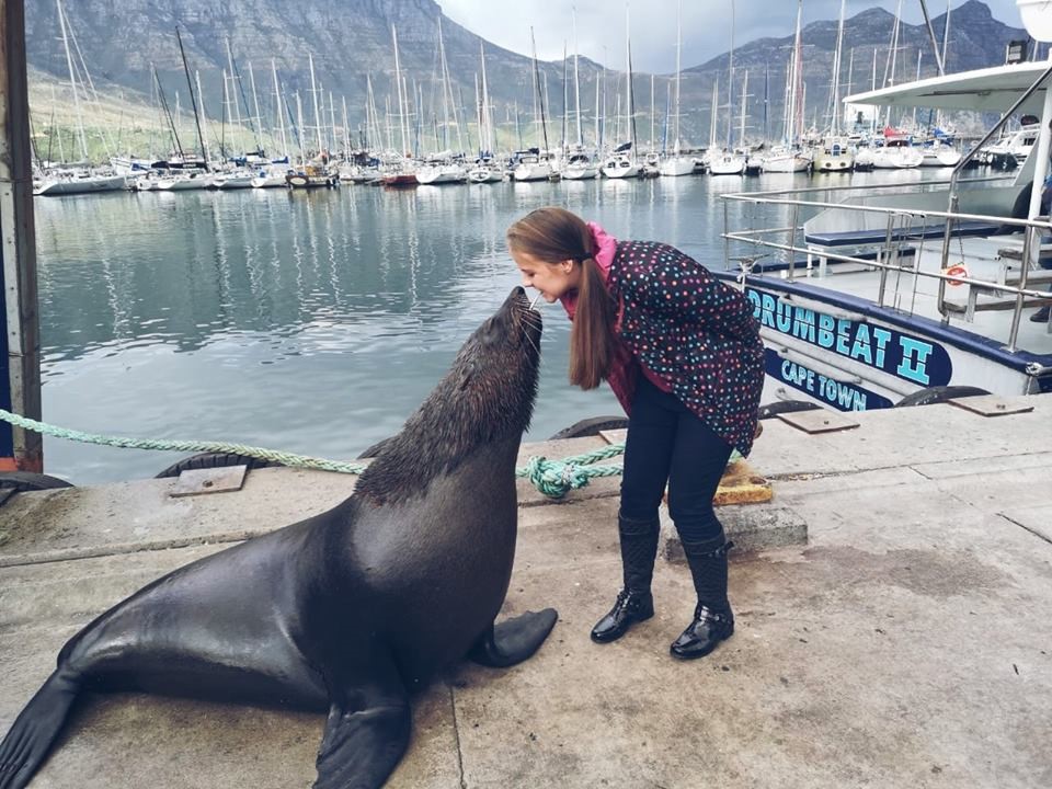
Figure 1) photo of tourist interacting with an adult male fur seal in Hout Bay
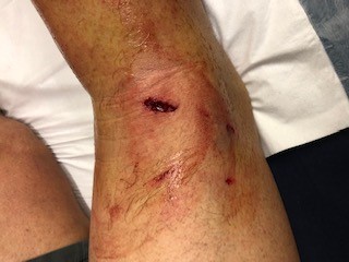
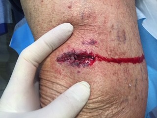
Figure 2, 3) Seal bite wounds that were treated in the Hout Bay facility
FIDSSA Members can earn CPD points by logging into the secure section of the website and visiting the MyCPD section.
Atlasville, Boksburg
South Africa
2022 © FIDSSA - All rights reserved • Website Terms of Use • Privacy Policy • Powered by E2
Admin login | Website login |
MYMEMBERSHIP®