ABOUT FIDSSA

Case of the Month

June 2018

Case courtesy of: Bernadett I. Gosnell, Sonal R. Verma, Virgilio J. Da Conceicao, Nithendra Manickchund, M. Yunus, S. Moosa, Department of Infectious Diseases, Nelson R. Mandela School of Medicine, and King Edward VIII Hospital, Durban

A 46-year-old female patient was seen at KEH VIII hospital.

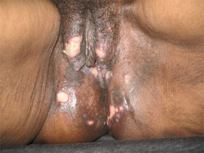
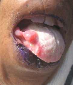
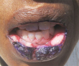
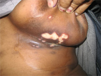
Presenting complaints: Non-healing ulcers of 4 months duration in the genital region, left infra-mammary region, right lateral tongue and lower lips. She also complained of weight loss, malaise and anorexia. She had no fever, no night sweats, no dysphagia, no odynophagia and no other gastrointestinal symptoms.

Past medical history: She was HIV-seropositive on first-line antiretroviral therapy (TDF/FTC/EFV) for the last seven years. Her most recent CD4 count was 3 cells/µL, viral load 29 056 RNA copies/ mL (4.46 log), suggesting that she had been failing treatment for a while. She was diagnosed with pulmonary tuberculosis four years ago and completed six months of appropriate treatment. She was known to be allergic to trimethoprim/ sulfamethoxazole

Social history: She did not smoke or drink alcohol. She was employed but had been on sick leave for the last two months.

Figure 1: Genital lesions

Figures 2-4: Ulcerative lesions on lip and inframammary region

Question 1: What are the commonest causes of genital ulcer disease in South Africa?

Answer to Q1

The majority of genital ulcers are caused by sexually transmitted infections (STIs). Non-infectious aetiologies should be considered once STIs have been ruled out. Of significance, genital ulcer disease (GUD) has been recognised as an important risk factor for not only acquiring HIV infection but also for transmitting HIV. A recent study from Johannesburg (Kularatne R. S. et al) reported that herpes simplex virus is the dominant cause of GUD, responsible for about 60% of all cases. This was followed by Treponema pallidum (3.9%), Chlamydia trachomatis (0.9%), Haemophilus ducreyi (0.5%) and mixed aetiology (0.8%). Of interest, no STI pathogen could be identified in 34.8% (Kularatne, Muller et al. 2018). This study also demonstrated a strong association between HIV seropositivity and GUD.

Question 2: How do genital ulcers impact on HIV disease?

Answer to Q2

HIV-associated immune dysregulation results in many diagnostic and therapeutic challenges. The control of STIs is important as this has long term public health implications. GUD has been recognised as a risk factor for acquisition for HIV. Several studies have demonstrated that GUD is more closely associated with HIV infection compared to other STIs. Several hypothesis have been suggested to explain this observation:

  • The ulcer itself serves as a portal of entry for HIV (Serwadda, Gray et al. 2003).
  • The associated mucosal inflammation recruits CD4+ T cells and other immune cells that serve as targets for HIV infection (Paz Bailey, Sternberg et al. 2010).
  • The ulcer may facilitate HIV transmission by increasing viral shedding from the genital area (Paz Bailey, Sternberg et al. 2010).
  • HIV disease with severe immunosuppression (CD4 count <50) is associated with an increased incidence of HSV GUD thus increasing the risk of HIV transmission. Genital ulcers secondary to HSV2 are associated with high HIV viral loads suggesting that HSV-2 infection may upregulate HIV replication (Serwadda, Gray et al. 2003).

Question 3: How should GUD be investigated once syndromic management fails?

Answer to Q3

A good history and clinical examination is very important to establish the cause of GUD. However, diagnostic dilemmas and therapeutic challenges always remain. The following investigations should be considered:
  • Swabs from the base of the genital ulcer for HSV culture or PCR. If vesicles or pustules are present, they should be unroofed and the base of the ulcer swabbed to obtain adequate cells for viral detection.
  • Rapid plasmin reagin (RPR) screen
  • Biopsy of the edge (of the freshest) ulcer
  • Smear, scraping or impression slides to stain for Haemophilus ducreyi, Klebsiella granulomatis (Calymmatobacterium granulomatis, Donovan bodies)
  • HIV test: If positive, institution of antiretroviral therapy is imperative.

Question 4: How do you confirm the diagnosis of HIV-associated genital ulcer disease?

Answer to Q4

This is a diagnosis of exclusion. Suspect this when patients have a low CD4 count and a high viral load or are on ineffective ART.

Question 5: What is the algorithm for management?

Answer to Q5

Flowchart: Adapted from ”Sexually Transmitted Infections Management Guidelines” 2015, South Africa

Effective antiretroviral therapy in this group of patients is critical for several reasons:

  • They are sexually active.
  • They are at high risk of transmitting HIV since they are practicing unprotected sex
  • They have gential lesions that increase their risk of transmission.

Case update

Our patient had a genital ulcer biopsy, which revealed chronic ulcerated skin with acute inflammatory exudate and granulation tissue. The stroma contained a dense mixed acute and chronic inflammatory cell infiltrate. No granulomas, acid fast bacilli, fungi or viral inclusions were identified.

HSV-PCR and CMV-PCR from samples taken from the base of the genital lesion, inframammary ulcer and lip lesions were all negative Biopsy of tongue biopsy was reported as non-specific with no evidence of inflammation or malignancy or granulomas Serology:
RPR was negative.
CMV IgG was positive and IgM negative.
EBV IgM was negative.
HSV was IgM negative.

ART regimen was changed to 3TC, AZT and LPV/r. Her CD4 count improved to 35 (from 3) cells/ µL and her viral load decreased to 164 copies/ mL within three months. Her genitial ulcers, and all extra-genital lesions resolved. We believe that these lesions were due to poorly managed HIV disease and with effective control of the HIV, all the lesions healed.

Earn CPD Points

CPD QUESTIONS

FIDSSA Members can earn CPD points by logging into the secure section of the website and visiting the MyCPD section.