ABOUT FIDSSA

Case of the Month

January 2017

Dr Ryan Aylward and Dr John Black

A 59 year old male from a residential area in Port Elizabeth was assessed in A&E with a short history of haematemesis and rash. He used alcohol regularly and was treated for pulmonary tuberculosis a few years prior, but was otherwise well. There was no significant travel or exposure history from the limited details available.

Pulse 125 bpm, BP 97/62mmHg and axillary temperature, 36.1°C. He had peripheral stigmata of chronic liver disease, a tender epigastrium, no ascites and the abdomen was not peritonitic. Purpura of the tip of the nose, trunk and digits were noted (figure 1). He had no features of infective endocarditis or meningitis.

Figure1

Chest x-ray revealed diffuse peri-hilar infiltrates and a right upper zone infiltrate (figure 3). There was no free air under the diaphragm. The peripheral smear showed intra- and extra-cellular bacilli (figure 2). The peripheral smear and laboratory results indicate a micro-angiopathic haemolytic anaemia (fragment count of 4.3%) with renal impairment. ADAMTS13 activity was normal (72%) and thrombotic thrombocytopenic purpura was therefore excluded. He was admitted to the ICU for acute haemodialysis and vasopressor support and was started on ceftriaxone. CT abdomen revealed renal cortical infarcts (figure 4) as well as an infarcted spleen of normal size.

Figure 1.

Figure 2.

Figure 3.

Figure 4.

Question 1: What infections are commonly associated with alcoholic liver disease?

Answer to Q1

The patient with alcoholic liver disease is prone to a variety of infections because of altered immune status and alterations in iron homeostasis. Social and behaviourial risk factors that may also predispose to infection include unfavorable living situations, poor oral and general hygiene, poor nutrition and risky sexual practices.

Specific organ systems involved include:
  • Pulmonary system – Poor dental hygiene with altered oral flora may harbor more virulent organisms including Klebsiella pneumoniae, Pseudomonas aeruginosa, and Enterobacter species. Altered mental status and increased risk of aspiration of oropharyngeal contents together with impaired neutrophilic mobilization and cytokine response predispose patients to community acquired pneumonia, lung abscesses and nosocomial pneumonia. Organisms such as Streptococcus anginosis, Streptococcus viridans, Staphylococcus aureus, Gram-negative bacilli, polymicrobial and anaerobic infections are often implicated. Tuberculosis is also more common in patients with alcoholic liver disease.
  • Cardiovascular - Pneumococcus, Bartonella quintana, and Capnocytophaga species are associated with endocarditis in this subset of patients.
  • Gastrointestinal – Reduced complement activation, impaired chemotaxis and phagocytosis, altered Kupffer cell functioning and shunting predispose to GI infections. Common infections include spontaneous bacterial peritonitis in those with ascites following development of portal hypertension as well as infected pseudocysts post pancreatitis. It is interesting to note that alcohol has some bactericidal qualities and there is an inverse correlation between alcohol consumption and the prevalence of H. pylori infections, with red wine having the most marked bactericidal effect.
  • Nervous system - Listeria monocytogenes is a well documented cause of CNS infections in alcoholics. In a review of central nervous system listeriosis, alcoholism/liver disease was the third most common predisposing factor for developing Listeria meningitis outside of the neonatal period and pregnancy following malignancy and transplantation. Listeria can cause meningitis, rhombo-encephalitis and brain abscesses.
Question 2 : What organisms are associated with a systemic sepsis syndrome in alcoholic liver disease?
Answer to Q2

Acute alcohol ingestion affects neutrophil adhesion, margination and migration. Chronic alcohol abuse affects cell mediated immunity at different levels. Lymphocyte proliferative response, monocyte production of pro-inflammatory cytokines, antibody-directed cellular cytotoxicity, and B-cell antibody-specific proliferation and production are all suppressed. Liver disease may be associated with functional hyposplenism and infections with encapsulated organisms such as Haemophilus, Pneumococcus and Meningococcus can occur. The acquisition of infections and risk of developing sepsis and septic shock are increased compared to non-alcoholic individuals with specific pathogens occurring more commonly: Vibrio vulnificus, Capnocytophaga canimorsus, Bartonella quintana, and Rickettsia species.

  • Vibrio vulnificus: This is a Gram-negative bacterium that causes severe wound and skin/soft tissue infections as well as sepsis. It is present in normal marine flora, commonly oysters, when water temperatures exceed 20°C. Consumption of contaminated raw oysters or exposure of a wound to contaminated salt or brackish water precedes wound infection and sepsis in most cases. The poor host cytokine response and the organisms’ ability to sequester iron ensure poor clearance and exponential growth. Bacteraemia can occur with no GIT symptoms. Patients present with an abrupt onset of chills and fever, followed by hypotension and the development of metastatic cutaneous lesions within 36 hours after onset of symptoms. Skin involvement starts with erythematous lesions that rapidly evolve to haemorrhagic bullae or vesicles and then to necrotic ulcerations. V. vulnificus bacteremia is fatal in more than 50% of patients, in whom this syndrome has been identified, including 100% of patients with hypotension. Patients with underlying liver disease and other chronic illnesses should avoid ingestion of raw oysters.
  • Capnocytophaga species are facultative anaerobic, gram-negative bacilli found in the oral cavity of humans and dogs. Patients without immune defects can present with a wide variety of syndromes, but asplenia and alcoholism have been identified as risk factors for septicemia caused by C. canimorsus. Other syndromes associated with bacteraemic patients include brain abscess, endocarditis, meningitis, acute respiratory distress syndrome, pneumonia, and wound necrosis.
  • Rickettsioses - Alcoholism is a risk factor for more serious outcomes of some of the rickettsial infections, notably:
  • Boutonneuse fever caused by Rickettsia conorii
  • Increased chronicity of Q fever caused by Coxiella burnetti
  • Rocky Mountain Spotted Fever (RMSF) caused by Rickettsia rickettsia
  • Epidemic typhus caused by Rickettsia prowazekii

Case continued

His clinical condition continued to deteriorate despite antibiotics. On further questioning his family volunteered that he had been bitten by a dog 2 weeks before presentation and on careful examination a small puncture wound was noted on his hand. Blood cultures were still negative.

Question 3 : What is the management of a dog or cat bite?
Answer to Q3

Management of a dog or cat bite includes the acute management of the injury and the risk of infection. Deep wounds pose a greater risk for infection. Prompt irrigation of the wound should be done with debridement of devitalized tissue and removal of foreign objects. Radiographs can be done to assess bony integrity and for the presence of foreign objects. Primary closure is generally not done and orthopaedic or plastic surgery evaluation may be required based on the extent and location of injury. Rabies prophylaxis should be considered on the basis of exposure risk and local epidemiological information. The need for tetanus vaccine or booster for the patient should be addressed. Re-examination should be scheduled at 48 hour post insult.

Preventive treatment with antibiotics are advised unless the bite is very superficial and easily cleansed. Because Pasteurella species are usually resistant to cloxacillin, clindamycin and erythromycin, these antibiotics are not recommended as monotherapy. Amoxicillin–clavulanic acid provides coverage against P. multocida, Capnocytophaga species, anaerobes and susceptible S. aureus. Alternatives include: doxycycline with metronidazole; clindamycin with a fluoroquinolone; or clindamycin plus co-trimoxazole in children. Established infections require admission to hospital for surgical debridement and drainage. Intraoperative cultures of necrotic/infected tissue should be obtained to ensure appropriate antibiotic choice and to allow de-escalation. Wound swabs are relatively uninformative due to the likelihood of culturing contaminating organisms.

Question 4: What additional pathogens should be considered in a patient with a dog bite?
Answer to Q4

In the setting of a dog bite, specific pathogens need to be considered. Most bite infections contain a mix of aerobes and anaerobes from both the skin of the patient and the mouth of the animal.
  • Aerobic organisms include: Pasteurella species, Streptococcus species, Staphylococcus species and Neisseria species
  • Anaerobic organisms include: Fusobacterium species, Bacteroides species, Porphyromonas species, Prevotella species, Capnocytophaga species.
Severe infections related to dog and cat bites can occur in about 20% of all cases, with complications developing more commonly in the hand (30–40%), where they are also the most likely site to develop rapidly spreading and permanently disabling damage. Cat bites are more likely to penetrate deeply resulting in deep abscesses and osteomyelitis more common. Patients presenting more than 8 hours after a bite often have infected wounds, with cat bites progressing to infection more rapidly than dog bites. Sepsis following a bite wound is often caused by those infected with C. canimorsus, P. multocida, Staphylococcus species (including MRSA), and Streptococcus species. Several other species, including Bacteroides, Fusobacterium, Neisseria and Prevotella might also produce bite-wound sepsis in immune suppressed individuals.
  • Pasteurella species are Gram-positive, facultative anaerobic, non-spore-forming bacilli. Virulence factors include capsular lipopolysaccharide, a cytotoxin, and iron acquisition proteins. P. multocida can be found as part of the normal flora of the upper respiratory tract of some mammals, particularly cats, with most human infections being caused by bites, although licks have also been associated with infection. Pasteurella species can cause serious infections, including necrotising fasciitis, septic arthritis, osteomyelitis and less commonly systemic sepsis and septic shock. Bacteraemia occurs in 25–50% of patients with pneumonia, meningitis and septic arthritis due to P. multocida and many have evidence of notable liver disease. In such cases, the mortality remains substantial at 25%. Rare complications include include peritonitis, meningitis and endocarditis. Like C. canimorsus, P. multocida is not susceptible to many oral antibiotics typically given for skin and soft tissue infections, including cloxacillin and clindamycin.

The history of a dog bite in an alcoholic with DIC prompted the search for Capnocytophaga canimorsus. Blood from still-incubated culture bottles was sent to the National Institute for Communicable Diseases in Johannesburg for 16S ribosomal RNA amplification. This PCR test confirmed Capnocytophaga canimorsus. Cases of meningitis and endocarditis have been confirmed with 16S PCR on CSF and valve tissue, respectively.

Question 5: Discuss the management of Capnocytophaga canimorsus
Answer to Q5

Bobo and Newton first described Capnocytophaga canimorsus in 1976. It is a Gram-negative bacillus found in normal oral flora in dogs and cats. Capnocytophaga canimorsus was previously known as DF-2 (Dysgonic fermenter-2) before being renamed by the Centre for Disease Control in 1989. The term ‘dysgonic’ was used to describe its slow growth in bacterial culture. In fact, the anti-coagulant Polyanethole-sulfonate used in culture medium has been implicated in inhibiting its growth.

C. canimorsus are capnophilic facultative anaerobic, fastidious Gram-negative rods. They are fusiform or filamentous gliding bacteria and are closely related to Fusobacterium and Bacteroides species. C. canimorsus requires large amounts of exogenous iron for growth. It grows slowly on blood (5% sheep blood in Columbia agar) or chocolate agar in 10% CO2. Media has to be incubated at 37.8C for at least 5 days. Colonies may not visible for 2–7 days on blood agar. Recently, the growth medium was optimized (Heart Infusion agar Difco with 5% sheep blood) and the bacteria were shown to grow in 2 days at 37.8C in 5% CO2.

C. canimorsus infection is associated with the absence of a pro-inflammatory response with low cytokine levels and is due to the fact that C. canimorsus does not interact with human Toll-like receptor 4 (TLR4) and is able to down regulate TLR4 and the pro-inflammatory signaling cascade. Not only is C. canimorsus resistant against phagocytosis and killing, it also blocks the killing of unrelated bacteria by macrophages.

Infection is most often transmitted through bites or contact with open wounds, but transmission has been described through contact with intact skin. Infection occurs after an incubation period of 1-7 days. Asplenic patients are at risk of more severe disease and alcohol abuse is another reported risk factor. No obvious risk factor can be identified in 40% of cases. Most infections occur in individuals older than 50 years.

The spectrum of C. canimorsus infections includes gangrene, sepsis, meningitis and endocarditis. Severe sepsis caused by C. canimorsus is extremely rare and mortality can range from 25–30% to as high as 60% in patients with septic shock. 60% of those who develop septic shock die within 30 days. A retrospective single centre Helsinki ICU cohort with a median age of 55.5 years found that 94% of patients with Capnocytophaga canimorsus were coagulapathic and 69% developed Acute Kidney Injury. Unexpectedly, patients with meningitis did relatively well.

Clinical manifestations:

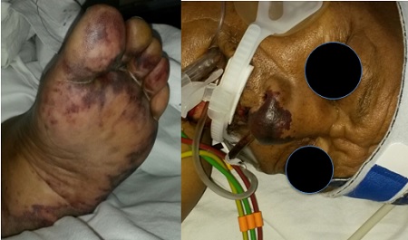
After an incubation period of 1–7 days, patients may experience an abrupt onset of malaise, abdominal pain that might mimic an acute abdomen, confusion, shortness of breath, and rapid progression to severe septic shock. On physical examination patients present with a petechial rash on the trunk, lower extremities, and mucous membranes. The original bite wound classically lacks inflammation. The rash might evolve from purpuric lesions to gangrene. The clinical manifestations of sepsis are secondary to a profound inflammatory response leading to endothelial dysfunction resulting in disseminated intravascular coagulation, acute respiratory distress and organ damage. Cases of fatal acute haemorrhagic adrenal insufficiency (Waterhouse–Friderichsen syndrome), gangrene and purpura fulminans may occur. Individuals aged more than 50 years are at greatest risk. Endocarditis and meningitis are difficult to differentiate from more classical causes other than from the underlying risk factors and exposure history.

Management:

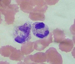
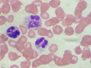
Diagnosis is usually based on clinical history because the bacteria are extremely difficult to grow. Up to 14 days of incubation might be necessary to detect growth on typical media. Peripheral blood smear and observation of abundant intracytoplasmic fusiform rods within neutrophils allows a presumptive diagnosis in the correct setting. Capnocytophaga species infection responds well to penicillin and β-lactam–β-lactamase inhibitor combinations. Other active agents include clindamycin, linezolid, tetracycline and carbapenems. Co-trimoxazole and aminoglycosides are not effective. Because of the relatively aggressive nature of infection and the difficulty in obtaining a laboratory diagnosis, therapy should be started as early as possible.

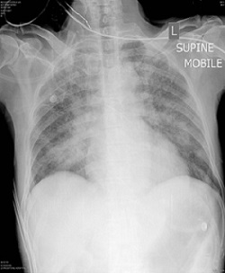
The patient subsequently developed hospital acquired pneumonia and antibiotics were escalated to Imipenem. The organism is sensitive to Carbapenems. He had a good clinical response and was eventually successfully extubated. He remained dialysis dependent for several weeks after which he was discharged with persistent renal dysfunction, but was not dialysis dependent.
Lessons Learnt

Even mild to moderate alcohol intake may affect immune responses. Factors such as lung micro-aspiration and social circumstances may further predispose alcoholics to infection. Specific organisms and sepsis syndromes need to be considered in patients who drink excessive alcohol. Of equal importance, patients with a history of a cat or dog bite may have been infected with unusual organisms like Pasteurella or Capnocytophaga and prompt wound care and prophylactic antibiotics should be given where appropriate. Fastidious organisms such as Capnocytophaga are difficult to grow on typical bacterial culture media and the possibility of such an infection in the right clinical setting should be communicated to the microbiology laboratory to ensure longer incubation time of cultures and an appropriate antimicrobial is used. The utility of the 16S PCR was also demonstrated in this case.
References:
  1. Gaastra W, Lipman L. Capnocytophaga canimorsus. Veterinary Microbiology 2010; 140: 339–346
  2. Kim A, Epstein M, Gautam-Goyal P, Thien-Ly D. Infections in Alcoholic Liver Disease. Clin Liver Dis 2012; 16: 783–803 http://dx.doi.org/10.1016/j.cld.2012.08.008
  3. Oehler R, Velez A, Mizrachi M, Lamarche J, Gompf S. Bite-related and septic syndromes caused by cats and dogs. Lancet Infect Dis 2009; 9: 439–47
  4. Jolivet-Gougeon A, Sixou J, Tamanai-Shacoori Z, Bonnaure-Mallet M. Antimicrobial treatment of Capnocytophaga infections. International Journal of Antimicrobial Agents 2007; 29: 367–373
  5. Butler T. Capnocytophaga canimorsus: an emerging cause of sepsis, meningitis, and post-splenectomy infection after dog bites. Eur J Clin Microbiol Infect Dis 2015; 34:1271–1280 DOI 10.1007/s10096-015-2360-7
  6. Pers C, Gahrn-Hansen B, Frederiksen W. Capnocytophaga canimorsus Septicemia in Denmark, 1982-1995: Review of 39 Cases. Clinical Infectious Diseases 1996;23:71-5
  7. Hastbacka J, Hynninen M, Kolho E. Capnocytophaga canimorsus bacteremia: clinical features and outcomes from a Helsinki ICU cohort. Acta Anaesthesiologica Scandinavica 2016; 60: 1437–1443 doi: 10.1111/aas.12752

Earn CPD Points

CPD QUESTIONS

FIDSSA Members can earn CPD points by logging into the secure section of the website and visiting the MyCPD section.