ABOUT FIDSSA

Case of the Month

April 2017

Dr SE Kubheka, M Archary and R Bobat, Department of Paediatrics Infectious Diseases, King Edward Hospital, University of KwaZulu-Natal

A 12-month-old boy was seen at a urban district hospital in KwaZulu-Natal with a 2-day history of fever, diarrhoea, irritability and abdominal distension. The stools were not blood-stained.

Three days prior to this admission his carer observed a single episode of a generalized tonic-clonic seizure associated with a fever. The infant was taken to the traditional healer where scarification of the body was performed. According to the family the infant improved over the next 2 days.

The rest of the history was unremarkable and age-appropriate vaccinations where up to date. Water source is piped water from a home tap.

On examination, his temperature was 38.5°C, pulse rate 140 beats per minute. He was well perfused but pale and irritable, no jaundice was noted.

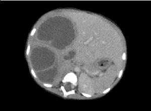
The only clinical abnormality of note was abdominal distention with a 5cm hepatomegaly without splenomegaly. Notably the neurological assessment was normal.

Initial investigations:

Question 1: What do you suspect and what would the next investigation be?

Answer to Q1:

With the hepatomegaly and changes in the right hemi diaphragm a liver abscess must be considered.

The next investigation needed is an ultrasound of the abdomen

Back to the case: The ultrasound in this child showed multiloculated heterogeneous masses in the right lobe of the liver, largest measuring 4cm x 5cm. The findings were suggestive of multiple liver abscesses.

Question 2: Which organisms can potentially be responsible and what empiric therapy would you initiate?
Answer to Q2:

(References as noted)

In cases of bacterial liver abscess polymicrobial infection is common.

Empiric therapy should consider specific risk factors and potential causative conditions that may indicate unique pathogens such as norcardia which can occur in patients with chronic granulomatous disease.

Community acquired cases of pyogenic liver abscess can be managed with a third generation cephalosporin. If E. histolytica is considered, metronidazole must be added, in addition to surgical interventions including percutaneous drainage.

Question 3: Which test would you order if a diagnosis of E. histolytica liver abscess is considered?

Answer to Q3:

Stool microscopy has a limited role to play in the diagnosis. Less than 30-40% of patients with amoebic liver abscess have concomitant intestinal amoebiasis, EIA for antibodies specific for E. histolytica is positive in approximately 95% of patients with extra-intestinal amoebiasis. These findings revert to negative in 6-12 months following eradication of infection however are also positive in intestinal infections and carriers

Back to the case:

The patient was initiated on Co-amoxiclav, Gentamycin and Metronidazole.

After 8 days of antibiotics the white cell count increased to 41 x 109/L, the hemoglobin dropped to 5.6g/dL, and there was no clinical improvement. The patient was transferred to the local referral centre.

At the referral centre the following tests were performed:

Entamoeba histolytica serology: IgG – positive (Ratio – 5.1)

CT scan of the abdomen:

Question 4: What is the likely diagnosis?

Discussion

Malaria and Entamoeba histolytica (E. histolytica) are the most common protozoal diseases worldwide.

E. histolytica most commonly presents with dysentery and liver abscess is its commonest extra-intestinal complication. In adults, for reasons that are poorly understood, extra-intestinal disease including liver abscess, is most common in men in the 4th and 5th decade of life, despite equal gender distribution of colonic amebic disease. Pulmonary and cerebral complications are rare. Defective cell mediated immunity (including HIV) is implicated in invasive disease.

Amoebic liver abscesses account for up to 34% of liver abscesses in children. Secondary infection is the most common complication of amoebic liver abscess (10–20%).

Presentation

Right upper quadrant pain

Low grade fever
Abdominal mass
Malaise
Diarrhoea

Risk Factors
Poor sanitation
Rural areas
High prevalence of protozoa

Complications
Rupture of the abscess occurs either into the pleura, lung, pericardium or the peritoneum. Abscesses located in the dome of the liver may rupture through the diaphragm and cause empyema, pleural effusion, bronchopleural fistula (4–7%). Those located in the inferior surface tend to rupture into the peritoneal cavity (7–11%). There have been reports of hepatogastric fistulas.

Differential diagnosis
Pyogenic abscess
Echinoccoccal disease
Tuberculosis
Congenital biliary and other congenital cysts and polycystic liver disease
Tumors and metastatic lesions

Investigations

Radiological
Ultrasound – commonly a unilocular heterogenous collection is seen in quadrant 4A, though it may occur anywhere in the liver.
CT abdomen – unilocular hypodense lesions are seen in the liver
MRI can also be used

Haematological
Full blood count – leucocytosis, anaemia
Presence of leucocytosis is not diagnostic as may be seen with pyogenic abscesses

Serology
Antibody detection tests
E histolytica IgG Elisa: Enzyme-linked immunosorbent assay for antibodies specific for E. histolytica is positive in approximately 99% of patients with extraintestinal amoebiasis findings revert to negative in 6-12 months following eradication of infection. In endemic areas a positive test is not helpful to confirm disease but a negative test, especially after the first week, can be helpful to exclude disease.

Antigen detection tests
E. histolytica antigen detection test has been shown to be more sensitive in detection of invasive extra-intestinal amoebic disease compared to antilectin antibody detection.
E. histolytica Real time PCR
It can detect the E histolytica DNA in blood, saliva and urine . Combined sensitivity of 97% has been reported if positive in urine and saliva in diagnosing amoebic liver abscess.

Treatment
Metronidazole alone given for two weeks has been shown to cure amoebic liver abscesses in up to 87% of patients. Some centres have performed percutaneous drainage in addition to metronidazole in 90% of their patients with amoebic liver abscesses.
Open abdominal surgery is necessary when the abscess has already ruptured or there is other abscesses in the abdomen.

Indications for percutaneous drainage
No clinical response in 48 – 72hrs
Abscess in the left lobe
Abscess more than 5 cm
Imminent rupture –thin rim or no parenchyma seen

Indications for open surgery
Failed percutaneous drainage, or abscess that is inaccessible to percutaneous drainage

Outcome:
We continued with the antibiotics and referred the patient for drainage of the abscess. Microbiological evaluation from the liver aspirate revealed scanty leucocytes and no bacteria were noted with a negative culture. Our patient had an uneventful recovery after percutaneous drainage

Lessons from the case
  • Importance of early surgical intervention in abscesses not responding to treatment within 72hrs.
  • Difficulty in conclusively diagnosing amoebic liver abscesses in an endemic area.
  • The atypical features of multiloculated abscess in amoebic liver abscess.
  • The empiric antibiotics probably treated the bacterial cause hence we did not culture anything in the pus.
Suggested reading
  1. Anshu Srivastava SKY. Identification of high-risk group and therapeutic options in children with liver abscess. European Journal of Pediatrics. 2012;171(1):33–41.
  2. Ba ID BA, Faye PM, Diouf FN, Sagna A. Particularities of liver abscesses in children in Senegal: Description of a series of 26 cases]. Archives de Pédiatrie. 2016;23(5):491–6.
  3. Cecilia Ximénez PM, Liliana Rojas. Novelties on amoebiasis: A neglected tropical disease. Journal of global infectious diseases 2011;3:166-74.
  4. Hanna RM DM, Badr SS, El-Betagy A. Percutaneous catheter drainage in drug-resistant amoebic liver abscess. Trop Med Int Health. 2000;5(8):578 -81.
  5. Haque R KM, Noor Z, Rahman SM, Mondal D, Alam F, Rahman I, Al Mahmood A, Ahmed N, Petri WA Jr. Diagnosis of amebic liver abscess and amebic colitis by detection of Entamoeba histolytica DNA in blood, urine, and saliva by a real-time PCR assay. Journal of clinical microbiology 2014;48(8):2798-801.
  6. Haque R MN, Ali IK, Alam K, Eubanks A, Lyerly D, Petri WA Jr. Diagnosis of Amebic Liver Abscess and Intestinal Infection with the TechLab Entamoeba histolytica II Antigen Detection and Antibody Tests. Journal of clinical microbiology 2000;38(9):3235-9.
  7. PL McGarr TM, SR Thomson. Amoebic liver abscess — results of a conservative management policy. S Afr Med J 2003;93:132-6.
  8. Rajak CL GS, Jain S, Chawla Y. Percutaneous treatment of liver abscesses: needle aspiration versus catheter drainage. AJR Am J Roentgeno. 1998;170(4):1035-9.
  9. Romano Ngui, Ishak S. Prevalence and Risk Factors of Intestinal Parasitism in Rural and Remote West Malaysia. PLoS Negl Trop Dis 5(3).
  10. Yun Liu J-yW. An Increasing Prominent Disease of Klebsiella pneumoniae Liver Abscess: Etiology, Diagnosis, and Treatment. Gastroenterology Research and Practice. 2013;2013(2013):12.

Earn CPD Points

CPD QUESTIONS

FIDSSA Members can earn CPD points by logging into the secure section of the website and visiting the MyCPD section.