ABOUT FIDSSA

Case of the Month

March 2016

Dr Bernadett Gosnell - King Edward VIII hospital/ NR Mandela School of Medicine, Durban
Ms Kavitha Naidoo – Dept of Infection Prevention and Control, University of Kwa-Zulu Natal, Durban
Prof MYS Moosa- King Edward VIII hospital/ NR Mandela School of Medicine, Durban
Prof AW Sturm - Dept of Infection Prevention and Control, University of Kwa-Zulu Natal, Durban

We describe a case of an HIV-infected, 27 year old female patient with a 5 year history of 1st line efavirenz-based antiretroviral therapy with long periods of defaulting treatment (CD4: 2 cells/µL and HIV viral load 80,000 copies/ mL). She presented with an enlarging, bleeding ulcer on her left introitus, which started 8 months prior with an indurated, painful nodule on the inner aspect of the left labia minora, which ulcerated spontaneously. No inguinal lymph nodes were palpable. She received benzathine penicillin 2.4 Mio IU by intramuscular injection as a single dose, erythromycin 500 mg orally six hourly for 1 week and acyclovir 400 mg orally eight hourly for one week without resolution of the lesion.

Question 1: What is the differential diagnosis for a genital ulcer?

Answer to Question 1

In sexually active young women, or in cases of sexual abuse, the differential diagnosis includes (most common in bold):
  • Herpes simplex virus HSV (most common, multiple vesicles progressing to pustules over 10-14 days)
  • Syphilis (painless ulcer)
  • Rarely- Lymphogranuloma venereum, Granuloma inguinale (Donovanosis) and Chancroid (ulcer is exquisitely painful and is associated with suppurative inguinal adenopathy)
The differential diagnosis of non-sexually transmitted vulvar ulcers is as follows (most common in bold)
  • Aphthous ulcers (synonyms include aphthosis, canker sores, Lipschutz ulcers, ulcus vulvae acutum)
  • Infectious
    • Herpes simplex virus (HSV) via autoinoculation
    • Ebstein Barr virus (EBV) (self-limited genital ulcers)
    • Cytomegalovirus (CMV) (rare)
    • Herpes zoster virus (VZV or HZV)
    • Group A Streptococcus (case reports)
    • Mycoplasma pneumoniae (case reports)
    • Molluscum contagiosum
  • Autoimmune (rare)
    • Crohn's disease (ulcers + GI symptoms)
    • Behçet's disease (Aphtous genital ulcers that last for weeks and heal with scarring)
    • Vaculitis (LUPUS)
    • Pemphigus and Pemphigoid (lesions may mimic lichen sclerosus with extensive scarring)
  • Drug reactions (rare)
    • Fixed drug eruptions (NAIDs, metronidazole, Acetaminophen [Paracetamol], Sulfonamides, Tetracycline, Phenytoin, oral contraceptives, Barbiturates, Phenolphthaten)
    • Stevens Johnson's syndrome/ toxic epidermal necrolysis
  • Other (rare)
    • Erosive Lichen Sclerosus
    • Hair removal folliculitis
    • Epidermolysis bullosa
    • Allergic contact dermatitis
Reference:
The Royal Children’s Hospital Melbourne. Clinical Practice Guidelines: Vulval Ulcers. Available at Visit website . retrieved on 22 february 2016

Further reading:
Bohl, TG. 2004. Vulvar ulcers and erosions-a dermatologist's viewpoint. Dermatol Ther. 17(1):55-67
Kirshen C., Edwards L. 2015. Noninfectious genital ulcers Semin Cutan Med Surg. 34(4):187-91

Case continued

In the case presented, the treatment did not improve the clinical picture and a second, most likely, seeded lesion developed.

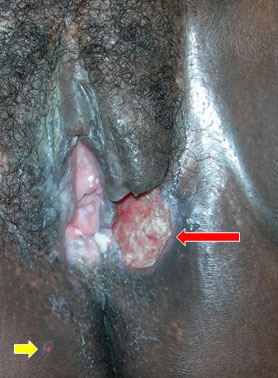
Image 1 (Published with permission of patient)

Red arrow: beefy red ulcer
Yellow arrow: early satellite lesion

Question 2: What is the most likely diagnosis when taking the clinical appearance into consideration?

Answer to Question 2

Donovanosis (granuloma inguinale), classic lesion of a beefy red ulcer

Answer to Question 3

  • Skin biopsy
  • Rapid plasmin reagin (RPR) screen
  • Pus swab for microscopy, culture and sensitivity (MCS)
  • Swab for Herpes simplex virus polymerase chain reaction (HSV PCR)
  • Smear, scraping or impression slides for staining techniques
Question 4: How can the diagnosis of Klebsiella granulomatis infection (Donovanosis) be confirmed?

Answer to Question 4

Donovanosis is a predominantly tropical cause of genital ulcer occurring chiefly in small endemic foci in all continents except Europe. Diagnosis requires the careful collection, staining and examination of smears or biopsies of characteristic genital, and occasionally, extragenital lesions for demonstration of the pathognomonic Donovan bodies within histiocytes. The causative organism is Klebsiella granulomatis (formerly Calymmatobacterium granulomatis), which is a Gram-negative, non-motile, pleomorphic bacterium that stains well with Giemsa, Wright, and silver stains. Donovan bodies are dark-staining (bipolar-staining) rod-shaped inclusion bodies seen in the cytoplasm of mononuclear phagocytes or histiocytes. Culture isolation of K granulomatis is difficult and impractical.

In the described case: Repeated pus swabs grew normal bowel and skin flora, a punch biopsy showed histological features of non-specific ulceration and impression slides sent to cytology confirmed an inflammatory process. The diagnosis of granuloma inguinale, Donovanosis, was made in the Department Infection prevention and Control, University of KwaZulu Natal, Durban on air-dried slides of scrapes from the edges and the base of the ulcer. Donovan bodies were identified with Giemsa (Rapi- diff) stain. The bacteria, Klebsiella granulomatis, are seen in a lysed white blood cell and stain blueish (image 2).

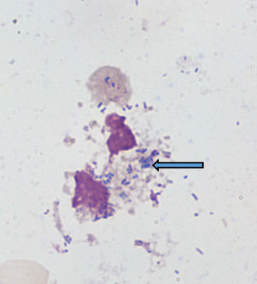
Image 2 with thanks from Prof AW Sturm’s laboratory, Durban, South Africa
(blue arrow pointing at a clump of Donovan bodies (Klebsiella granulomatis))

Reference
Richens, J. 1991. The diagnosis and treatment of donovanosis (granuloma inguinale). Genitourin Med 67(6):441-452

Question 5: How would you treat this patient?

Answer to Question 5

Several antimicrobial regimens have been effective, but only a limited number of controlled trials have been published (O'Farrell. 2002). Treatment has been shown to halt progression of lesions, and healing typically proceeds inward from the ulcer margins; prolonged therapy is usually required to permit granulation and re-epithelialization of the ulcers. Relapse can occur 6–18 months after apparently effective therapy. The Centers for Disease Control and Prevention (CDC) STD treatment guidelines should be followed.

Recommended Regimen

Azithromycin 1 g orally once per week or 500 mg daily for at least 3 weeks and until all lesions have completely healed

Alternative Regimens

Doxycycline 100 mg orally twice a day for at least 3 weeks and until all lesions have completely healed
OR Ciprofloxacin 750 mg orally twice a day for at least 3 weeks and until all lesions have completely healed
OR Erythromycin base 500 mg orally four times a day for at least 3 weeks and until all lesions have completely healed>
OR Trimethoprim-sulfamethoxazole one double-strength (160 mg/800 mg) tablet orally twice a day for at least 3 weeks and until all lesions have completely healed.

References
O'Farrell, N. 2002. Donovanosis. Sex Transm Infect 78:452-7
Centers for Disease Control and Prevention (CDC). 2015. Sexually Transmitted Diseases Treatment Guidelines. Available from www.cdc.gov/std/tg2015/donovanosis.htm . accessed on 22 February 2016

Outcome of the case:

A 6 week course of azithromycin 500 mg orally on alternate days led to complete resolution of both lesions. Her ARVs were changed to second line therapy consisting of AZT/3TC and LPV/r. At 6 weeks: CD4 13 cells/µL, VL < 150 copies.

Earn CPD Points

CPD QUESTIONS

FIDSSA Members can earn CPD points by logging into the secure section of the website and visiting the MyCPD section.