ABOUT FIDSSA

Case of the Month

September 2015

Frans Radebe – Centre for HIV & STIs, NICD/NHLS

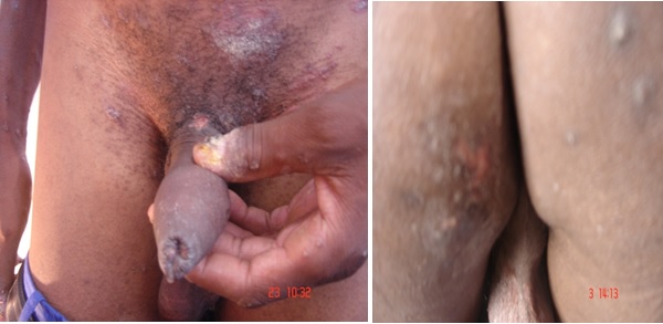
A 29-year man presented at a dedicated Sexually Transmitted Infections (STIs) clinic in Johannesburg, complaining of a mucocutaneous, florid eruption on the external extremities involving fingers, arms, buttocks, neck, penile shaft and face for 3 weeks. Prior to that, he noticed a painless, solid ulcer which was in the process of healing without receiving treatment. He also complained about headache, body weakness and discomfort due to icthiness of the rash. He was uncircumcised and had phimosis of the penis. No history of anal intercourse was reported.

On physical examination, the diffuse mucocutaneous rash was evident, in addition to patchy alopecia, and non-tender, regional lymphadenopathy.

Genital swabs were taken after the ulcer was scraped to expose the lesion. Swabs were also collected from areas of rash on the proximal extremities, and laboratory testing by a Multiplex PCR, for HSV-2, Treponema pallidum, Haemophilus ducreyi and Chlamydia trachomatis L1-3 strains was undertaken. Blood sample was collected for serological testing for syphilis and HIV rapid testing.

His record card indicated that he had been treated for syphilis three years previously and had tested HIV-seropositive.

He was treated syndromically for genital ulcer syndrome with Benzathine benzyl penicillin IM, 2.4 MU and since his HIV status was known, Acyclovir, oral 400mg 8 hourly for 7 days was added. He was asked to return to the clinic after a week for further assessment with the laboratory report for PCR and serology.

Questions

  • What is the likely diagnosis and describe its clinical presentation?
  • What are the common differential diagnoses?
  • What role does laboratory testing play in terms of diagnosis and treatment?
  • How would you treat the case?

Q1: Diagnosis and clinical features of the infection

Syphilis infection passes through a series of frequently overlapping stages which need to be taken into consideration when making a diagnosis. With the recent increase in syphilis infection among certain key populations, overall climb in rates in certain geographic areas of the world and the HIV epidemic, clinical and serological diagnosis of syphilis may present with many challenges to the clinician. Attention to the time course of symptoms and signs is required for proper disease staging namely primary, secondary, latent (early and late) and tertiary syphilis. In primary infection, patients typically develop a skin lesion (chancre) on the site of inoculation within 3 weeks after exposure to an infected individual. The chancres frequently go unnoticed particularly among women or men who have sex with men who may not be able to see vaginal or anal lesions. The lesions are classically nontender, indurated, and nonpurulent. More than 70% of patients present with rash, which may be macular, pink or red, distributed on shoulders, arms, chest, back and other external extremities. If untreated, the macules can become maculopapular affecting the hands and soles. The rash of secondary syphilis can be pustular, follicular but are hardly ever vesicular. The secondary infection is a systemic process that can infect any organ of the body. It resolves without treatment but recurrences are common and the patient develops late sequelae following many years of silent infection.
Q2: Differential diagnosis

A syphilitic ulcer should be differentiated from the soft chancre of chancroid caused by Haemophilus ducreyi on clinical grounds; chancroid ulcers are tender with jagged border and have yellow exudates. A smaller chancre may be due to Lymphogranuloma venereum (LGV). A lesion resembling a chancre may also be caused by trauma to the penis or drug eruption. The syphilitic lesions may be similar to lesions caused by herpes simplex infections. Co-infections are common and secondary bacterial infections may complicate the diagnosis. As the manifestations of syphilis or advanced syphilis are sometimes non-specific, a high index of suspicion must be kept regarding the possible diagnosis of the disease. Attention to the time course of symptoms is required for thorough sexual and social history such as the number of sexual partners, condom use, history of STIs in the patient and partners, drug use and previous exposure to blood use or its products. Differential diagnosis is crucial in stages of infection as other signs and symptoms may vastly differ especially in secondary syphilis.
Q3: The role of laboratory testing for diagnosis and treatment.

Treponema pallidum cannot be cultured in vitro and the organism is too small to be seen under the light microscope. However, Dark-field microscopy is essential in evaluating moist cutaneous chancre lesions of primary syphilis or the condyloma lata of secondary infection. Alternatively, direct immunofluorescence staining of fixed smears by direct fluorescent antibody Treponema pallidum test can be used.

Serologic testing is considered the standard method of detection for all stages of syphilis. However, serologic tests cannot be used to differentiate the different species of the treponema family.

In suspected acquired syphilis, traditional testing is performed with a sensitive nontreponemal screening test; venereal disease research laboratory (VDRL) or rapid plasma reagin (RPR). Coexisting conditions of TB and malaria among others may reduce the tests specificity. VDRL becomes positive 1-2 weeks after chancre formation and both tests become nonreactive with time after treatment or can persist for life in some patients and can be quantified. With the possibility of false positives, confirmation of any reactive results by a treponemal test is warranted. Confirmatory tests may include fluorescent treponemal antibody-absorption (FTA-ABS), T.pallidum hemagglutination (TPHA), T.pallidum particle agglutination (TPPA), EIA IgG and IgM for treponemal. Diagnosis of neurosyphilis is made by a VDRL-CSF test which is highly specific with a low sensitivity, thus a need for a combination of reactive serologic tests results, CSF cell count and CSF protein measurement.

In Congenital syphilis, a positive VDRL or FTA-ABS can be confirmed by 19S IgM FTA-ABS since maternal IgM is not passed to the fetus in utero or a Captia-Syphilis –M enzyme-linked immunosorbent assay. Due to the prozone phenomenon false negative results are common in persons with very high titer RPR or VDRL who may become positive with serum dilution. HIV infection may also cause false-negative results in both nontreponemal and treponemal tests. Contrary to the norm, some patients with primary or secondary syphilis may have persistent positive nontreponemal test results 36 months after treatment.

Recently, reverse sequence screening test have been proposed in order to reduce time, labor and costs where screening is by a treponemal ELISA and reactive sera are confirmed with a nontreponemal test.
Outcome of the case

As arranged with the patient during the first visit, he came back for assessment after a week. The rash had improved, but pigmentation was still very visible in most of the extremities. He was feeling much stronger and the headache was not infrequent. The ulcer was healing very well and he was positive for Treponema pallidum and HSV-2 BY MPCR. Syphilis serology was still positive at a titre of 1:32 and another blood sample was collected to monitor the treatment progress and Benzathine penicillin 2.4 MU injection was increased for once weekly for 3 weeks and it was dissolved in 6ml l% lidocaine without epinephrine to ease the pain.

Earn CPD Points

CPD QUESTIONS

FIDSSA Members can earn CPD points by logging into the secure section of the website and visiting the MyCPD section.