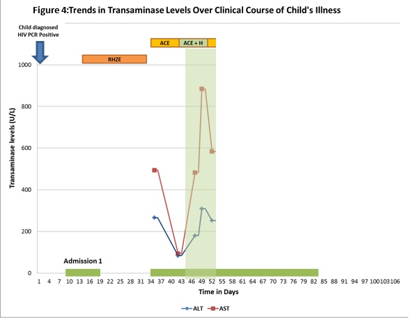
Title: A Little Liver and a Big TB problem
David P Moore - Department of Paediatrics and Child Health, Chris Hani Baragwanath Academic Hospital and University of the Witwatersrand david.moore@wits.ac.za
A 6-week old female infant, newly diagnosed with HIV, was started on appropriate doses of anti-tuberculosis treatment on day five of her sentinel hospital admission with pneumonia, in view of poor weight gain (severe acute malnutrition: weight 3.1 kg (weight-for-age Z-score: -3.29 SD), a ‘suggestive chest X-ray’ and splenic microabscesses visualised on the abdominal ultrasound (Figure 1).
Figure 1: Abdominal ultrasound from first hospital admission
Ultrasound of the spleen demonstrates a diffuse coarse echogenicity with multiple hypoechoic lesions highly suggestive of microabscesses. The radiologic differential for microabscesses in the spleen includes disseminated TB, non-tuberculous mycobacterial infection (Mycobacterium avium complex), and fungal infections.
Her CD4 count was 53 cells/mm3 (2.02%). The baseline viral load was 4,170,560 RNA copies/mL. There was no known TB contact.
She was treated for community-acquired pneumonia, and responded well to therapy. She was discharged four days later in good health, with a plan to initiate her onto antiretroviral therapy (ART) in 4 weeks, once stabilised on anti-TB treatment.
Question 1: According to the World Health Organization, what clinical stage of HIV does this child fall into?
Question 2: What investigations can be sent to confirm a microbiological diagnosis of tuberculosis in this infant?
Question 3: What anti-tuberculosis regimen would you use in such a child?
Question 4: What doses of first-line anti-tuberculosis drugs are recommended for children, according to the South African TB Guidelines?
Question 5: What additional medication is warranted in such a child, and why?
Frontal chest X-ray demonstrates reticular pattern in the right middle zone, with some obscuration of the right heart border. The child is rotated in this X-ray, but there are no obvious features to suggest hilar adenopathy, i.e. no lobulated, dense masses filling the hilar points
Question 6: Why would a liver function test be warranted in this infant, despite the fact that she was not clinically jaundiced?
In the repeat ultrasound, the liver appears homogenous with no focal areas of increased or decreased echogenicity. There are no obvious calcifications. There is normal colour flow on Doppler ultrasound of the portal and hepatic veins. Resistive index to flow in these vessels was not calculated. The splenic microabscesses are less prominent.
Question 7: Are these transaminase levels a cause for concern, and if so what differential diagnosis would you consider as a cause for this clinical picture?
ACE: amikacin, ciprofloxacin, ethambutol | AST: aspartate transaminase |
ACE + H: amikacin, ciprofloxacin, ethambutol and isoniazid | RHZE: rifampicin, isoniazid, pyrazinamide and ethambutol |
ALT: alanine transaminase |
3TC: lamivudine | AME +R: amikacin, moxifloxacin, ethambutol and higher-dose rifampicin |
ABC: abacavir | ART: antiretroviral therapy |
ACE: amikacin, ciprofloxacin, ethambutol | AST: aspartate transaminase |
ACE + H: amikacin, ciprofloxacin, ethambutol and isoniazid | LPV/r: lopinavir and ritonavir |
ALT: alanine transaminase | MERZ: moxifloxacin, ethambutol, rifampicin and pyrazinamide |
AME: amikacin, moxifloxacin, ethambutol | RHZE: rifampicin, isoniazid, pyrazinamide and ethambutol |
AME +r: amikacin, moxifloxacin, ethambutol and low-dose rifampicin |
Discussion:
Toxicity to first-line anti-TB drugs is uncommon in clinical paediatrics, however it may be devastating if not managed appropriately [5]. Although isoniazid, rifampicin and pyrazinamide all have the potential for causing hepatocyte damage, isoniazid is the most frequently implicated agent [6]. Isoniazid is metabolised by N-acetyltransferase in the liver, and young children tend to metabolise the drug more efficiently than do older children and adults [7]. Recommended doses of isoniazid in the South African Paediatric TB Guidelines (10-15 mg/kg/day) and revised World Health Organization (WHO) Guidelines (10 mg/kg/day) is higher than those previously recommended in paediatric treatment guidance documents ( ̴5 mg/kg/day) [8].
It is expedient to do baseline liver function tests (ALT, AST) in young HIV-infected children who are on anti-TB treatment, in whom we are soon to initiate ART [9]. No baseline liver functions were done in the child reported here, and the hepatitic picture would have been missed entirely if the test was not conducted at the second hospital admission.
A positive TB blood culture in this child is reassuring of the fact that continuation of anti-TB therapy is warranted in this child. Duration of anti-TB therapy is debatable, but as isoniazid cannot be used with safety, at least 12 months of treatment is justified.
Acknowledgements
Thank-you to Dr Pedro da Silva (Braamfontein TB Laboratory) for assistance in interpreting the TB blood culture result), and Dr Nasreen Mahomed (Respiratory & Meningeal Pathogens Research Unit, ) for expertise in interpreting the radiological investigations done in this case. Ethics permissions for online publication of this case were obtained from the Chris Hani Baragwanath Medical Advisory Committee and the University of the Witwatersrand Human Research Ethics Committee (Medical)
References:
FIDSSA Members can earn CPD points by logging into the secure section of the website and visiting the MyCPD section.
P.O. Box 16011,
Atlasville,
1465
South Africa
2022 © FIDSSA - All rights reserved • Website Terms of Use • Privacy Policy • Powered by E2
Admin login | Website login |
MYMEMBERSHIP®