Dr Jeremy Nel, Dept of Infectious Diseases, Helen Joseph Hospital
A 28-year-old man presented at a local district hospital complaining of a chronic cough (> 1 month duration) with associated right-sided pleuritic chest pain, anorexia and weight loss. A chest X-ray showed a right-sided pleural effusion. An intercostal drain was inserted, and the pleural fluid taken before antibiotics were commenced, showed:
Pleural fluid cytology revealed no malignant or mesothelial cells. A second chest X-ray, taken after the intercostal drain insertion, additionally showed a right pneumothorax, revealed after drainage of several litres of purulent fluid.
His blood tests on admission were as follows:
Question 1: What is the differential diagnosis of a chronic exudative pleural effusion, considering the negative Gram stain and routine bacterial culture?
Answer to Q1
A chronic, exudative pleural effusion with a negative Gram stain and routine bacterial culture, should prompt the following differential diagnosis, amongst other things:
Note: some of the above may be cultured on routine bacterial culture (e.g. Nocardia) although the sensitivity for their detection by this method is low, and therefore they should remain considerations at this stage.
A CT scan of the chest revealed a significant residual right pneumothorax (despite the intercostal drain), and collapse/consolidation of all 3 lobes of the right lung, with mediastinal shift to the left:
An alert clinician noted that the fluid, while grossly purulent, had an unusual brown-orange colour (see picture below) and was odourless.
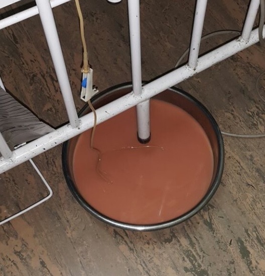
Question 2: An ‘empyema’ that looks like this is virtually pathognomonic for which diagnosis?
Answer to Q2
The pleural fluid shown has the appearance of “anchovy paste”, and this is virtually pathognomonic for amoebiasis.
The “anchovy paste” appearance is the result of a combination of necrotic and apoptotic material, and haemolysis of red cells that have bled into the cavity.
Question 3: Which is the most appropriate diagnostic test that can be performed to confirm this diagnosis?
Answer to Q3
The most appropriate diagnostic test at this stage would be serology for amoebiasis.
More than 95% of both amoebic liver abscesses and thoracic amoebiasis develop detectable antibodies. Such testing may be performed by indirect haemagglutination assays (IHA) or ELISA assays .
Microscopy may demonstrate trophozoites from aspirated material, but sensitivity is poor.
Antigen testing and molecular diagnostics (PCR) on aspirated material are both highly sensitive and, especially in the case of PCR, highly specific. However, they are expensive and generally limited to reference laboratories in Southern Africa. In the context of this case, they would be unlikely to add anything to serology, which is much cheaper and more readily available.
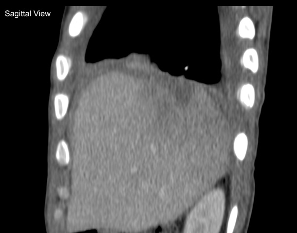
Question 4: Primary amoebic empyemas are very rare. Where else should one look in the body to see if there is another (primary) focus of infection?
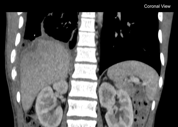
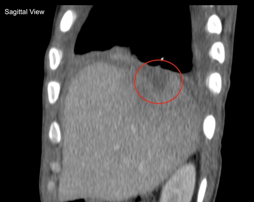
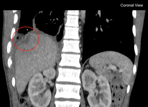
Answer to Q4The abdomen should be scanned to look for a ruptured amoebic liver abscess. Almost all amoebic empyemas are the result of such an event1.
Although both an abdominal ultrasound and a contrasted abdominal CT scan were initially reported as normal, alert infectious diseases doctors insisted on a review of the scans.
Question 5: In the cuts from the abdominal CT scan above, what do you see?
Answer to Q5
The subtle defects circled below show a ruptured, healing abscess, situated immediately below the diaphragm. This is precisely the location where abscesses that rupture into the pleural space are most likely to be found.
Question 6: How should amoebic liver abscesses and amoebic empyemas be treated?
Answer to Q6
In general, extra-intestinal amoebiasis requires both3:
Surgical drainage is generally not required for amoebic abscesses as an excellent response to therapy can be anticipated2.
Our patient required decortication of the lung, as it failed to re-expand despite both the insertion of an intercostal drain & appropriately-provided negative suction pressure to the pleura. He made a full recovery after the combined medical and surgical therapy however.
Lessons learnedReferences
FIDSSA Members can earn CPD points by logging into the secure section of the website and visiting the MyCPD section.
Atlasville, Boksburg
South Africa
2022 © FIDSSA - All rights reserved • Website Terms of Use • Privacy Policy • Powered by E2
Admin login | Website login |
MYMEMBERSHIP®