
Dr David Stead – IDSSA (thanks to Dr Nelesh Govender & Dr Sean Wasserman for their assistance)
A 16 year old girl with chronic granulomatous disease diagnosed in early childhood, on cotrimoxazole and itraconazole as primary prophylaxis, presented to a private physician with fever, neck pain, dysphagia, weight loss, and mild weakness of her hands over a few weeks. On examination there was no weakness or spasticity of her upper limbs. White cell count was normal, but her C-reactive protein was elevated at 123. Lumbar puncture was normal. An MRI spine was performed:
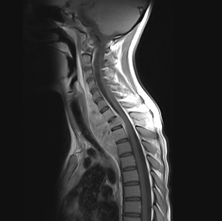
This was reported as enhancement of C7-T2 with volume loss of T1 vertebral body. An associated enhancing paravertebral mass without cord compression, with a large prevertebral component was noted. There were also enhancing nodules in the left lung apex and hilar region (no CXR available). Surgical evacuation of the prevertebral mass was performed.
Question 1: What infectious aetiologies would you consider?
Answer to Q1
Chronic granulomatous disease (CGD) is a rare primary immune deficiency affecting 1 in 200 000 live births. It is characterized by phagocyte dysfunction due to genetic mutations encoding phagocyte nicotinamide adenine dinucleotide phosphate (NADPH) oxidase. The reduced microbicidal oxygen radical production renders sufferers susceptible to severe bacterial and fungal infections. Some CGD patients manifest with auto-immune or granulomatous complications requiring immune-suppression. Most cases are diagnosed in early childhood. More than two thirds are X-linked recessive, the remainder autosomal recessive.1
The majority of infections in a US cohort of 250 CGD cases were due to 4 bacteria: Staphylococcus aureus, Serratia marcescens, Burkholderia cepacia complex, and Nocardia species, as well as species of the fungus Aspergillus. The lung was the most frequently affected organ, and 40% of culture positive organisms were Aspergillus species. Liver abcesses, skin infections, and lymph nodes are other commonly affected sites.2
A recent review of 46 published cases of Aspergillus osteomyelitis in CGD reported 50% due to A. fumigatus, and 43% due to A. nidus. There was a significantly higher mortality in the A.nidus (55%) vs A.fumigatus (13%) groups (p=0.008). Most cases are due to contiguous spread from a pulmonary focus, with the vertebra most commonly involved.3
In summary, the most likely organisms in this case would be: Aspergillus species, Serratia marcescens, Staphylococcus aureus, Nocardia, and in a high TB prevalence area, Mycobacterium tuberculosis.
The pus cultured Aspergillus spp, and she was started on voriconazole 100mg twice daily. She had a good initial clinical response and a follow up CRP after 5 weeks was 8. There was further wedge compression of T1 on follow up imaging, and the specialist team elected for prolonged voriconazole therapy.
After 12 months on voriconazole therapy, she developed progressive neck pain, dysphagia and subtle left hand weakness. Clinically she had 4+/5 power in C7/T1 root motor groups of her left hand. She was otherwise well. A repeat MRI spine was performed:
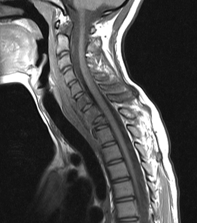
Report: Abnormal marrow signal in bodies and posterior elements of C6 - T1 with complete collapse of T1. Prevertebral increased signal in the soft tissues of the neck suggesting early myositis/cellulitis rather than true abscess formation, involving neural foramina at multiple levels.
Question 2: What are possible explanations for the apparent disease progression after 12 months of therapy?
Answer to Q2
1. Non-compliance to the voriconazole:
After careful counselling, she admitted to taking approximately 50% of the prescribed voriconazole therapy. This was partly due to side effects, mostly red - green visual disturbance.
2.Inadequate dose/levels:
The recommended dose of voriconazole is a 6mg/kg IVI loading dose for 24 hours, then 4mg/kg 12 hourly IVI. The usual oral dose is 200mg bd, but 4mg/kg orally bd is also used. She weighed 45 kg, thus requiring a minimum of 180mg bd (originally underdosed on 100mg bd).
Voriconazole has non-linear pharmacokinetics and its dose-response relationship exhibits wide interpatient variability. The therapeutic index is narrow, and serum concentrations are significantly influenced by a broad range of drug-drug interactions. The cyP2c19 isoenzyme, for which voriconazole is an important substrate, displays frequent genetic polymorphisms. Poor metabolizers can have up to 4 times higher serum voriconazole concentrations than extensive metabolizers4. In light of unpredictable levels, routine therapeutic monitoring is recommended, especially in the context of a poor treatment response5.
After increasing her dose to 200mg bd, voriconazole trough level was measured at 2.8mg/l (1-6mg/l recommended).
3. Voriconazole resistance:
The local prevalence of voriconazole resistance for Aspergillus is unknown. Elsewhere, resistance has been shown to emerge where patients are treated with long-term azoles, but also in the azole naïve. The prevalence of resistance in clinical isolates reported elsewhere ranges from 1% to 18%6
Question 3: How would you manage her?
Answer to Q3
Induction therapy: She was given 2 weeks of IV amphotericin B for suspected active Aspergillus disease.
Consolidation therapy: Voriconazole is the recommended empirc first line treatment for Aspergillus infection5. In the absence of microbiological evidence for resistance, and with the history of poor compliance, we elected to continue with this, but at 200mg bd. Trough levels were found to be therapeutic at this increased dose.
Alternative salvage options include amphotericin B, caspofungin and posaconazole (although a small rate of cross-resistance to voriconazole occurs) or a combination of the latter two. There is little evidence for these strategies.6
Secondary prophylaxis: Lifelong voriconazole would be recommended in this case.
Immunotherapy: Immunotherapy appears to be an important adjunct in the treatment of invasive aspergillosis in CGD patients. Both IFN-γ and haematopoietic cytokines, such as G-CSF and GM-CSF, have been used to facilitate the cure of aspergillosis in CGD patients.3 Further discussion of this is beyond the scope of this case.
The IDSA guideline on Aspergillus osteomyelitis states: “Surgical resection of devitalized bone and cartilage is important for curative intent.” Approximately two thirds of the previously mentioned 46 aspergillus osteomyelitis in CGD underwent surgery, but it was associated with a non- significant trend to poorer outcomes.3 While a repeat biopsy and tissue culture would have been greatly informative, and debridement of the T1 vertebral body potentially aided sterilization, the orthopaedic surgeons felt the risks outweighed the benefits in this case.
Our patient had a good clinical response to the medical therapy above, regaining full power by the end of the Amphotericin B therapy, and remaining largely asymptomatic on the increased voriconazole dose. She is planned for repeat MRI and voriconazole level, and is receiving adherence counselling.
References:
FIDSSA Members can earn CPD points by logging into the secure section of the website and visiting the MyCPD section.
Atlasville, Boksburg
South Africa
2022 © FIDSSA - All rights reserved • Website Terms of Use • Privacy Policy • Powered by E2
Admin login | Website login |
MYMEMBERSHIP®