
Carin Erasmus
Infection Control Society of Southern Africa (ICSSA)
An infant who had been discharged from the neo-natal intensive care unit (NNICU) was re-admitted to the paediatric unit, within 21 days, with respiratory distress. He was one of a twin and born prematurely (Case 2,Table 1). Five days after his re-admission, the infant had to be transferred back to the NNICU. He was unstable and required oxygen, and the radiological findings were compatible with Pneumocystis pneumonia (PCP). An induced sputum sample was positive by PCR for Pneumocystis jiroveci.
The hospital was informed by the Paediatrician that another premature infant (case 1), had been re-admitted to a NNICU in another hospital with PCP. This led to heightened vigilance and testing of neonates in the unit. Over the next 2 months, a cluster of 7 premature infants were diagnosed with PCP (Table 1) and from July - October, another cluster of 4 cases occurred.
Of the 11 infants, 9 were discharged apparently well and re-admitted to a health care facility, requiring additional oxygen within time periods of 11 to 109 days after birth. The remaining 2 infants remained in the NNICU for 62 and 100 days respectively before being diagnosed with PCP in the Unit after they too, required supplemental oxygen.
Question 1: What is the significance to neonates of P. jiroveci colonisation in the human host?
Answer to Q1
Pneumocystis jiroveci, previously known as Pneumocystis carinii, has long been recognized as an opportunistic organism which causes infections in the lower respiratory tract of humans. Individuals with a weakened immune system are most commonly affected. Pneumocystis colonisation is defined when Pneumocystis detection is present in an individual without signs and symptoms of Pneumocystis pneumonia.
Post mortem studies as well as screening of respiratory samples indicate that P.jiroveci can be detected in the lungs and airways of both healthy and immuno-compromised individuals, who are asymptomatic. Distinct species of Pneumocystis are strictly linked to a specific mammalian host. Discoveries have also led to the prevailing insight that the human population is the only reservoir for P. jiroveci.
The significance of Pneumocystis colonization is not yet fully understood, although colonization may be a prelude to clinical disease and is a resevoir for transmission. Vargas et al, demonstrated the ability of Pneumocystis to replicate in the lungs of immunocompetent hosts. Thus, transmission of the pathogen among medical personnel and patients is likely and transmission from mother to the neonate remains possible.
Question 2: Which neonates are most likely to be pre-disposed to PCP?
Answer to Q2
Pneumocystis carini forma specialis hominis affects severely ill and immature neonates and epidemics in NICCUs have been described. Neonates’ cellular and humoral responses are compromised. Surfactant deficiency and coexisting infection may heighten predispostion, as may protracted mechanical ventilation, antibiotics and steroids.
When the lungs contain normal amounts of surfactant, growth of Pneumocystis in the host is inhibited. Alterations in the quantity of surfactant contribute to PCP. In a study states of congenital deficiency of surfactant 90.5% of neonates in the PCP group had surfactant deficienty, compared to only 43.2% of the control group (Kordek). Further studies describing neonates with PCP, showed documented low birth weight and more common need for steroids to accelerate lung maturation.
The significance of Pneumocystis colonization is not yet fully understood, although colonization may be a prelude to clinical disease and is a resevoir for transmission. Vargas et al, demonstrated the ability of Pneumocystis to replicate in the lungs of immunocompetent hosts. Thus, transmission of the pathogen among medical personnel and patients is likely and transmission from mother to the neonate remains possible.
Question 3: Should premature infants receive trimethoprim-sulfamethoxazole prophylaxis to prevent Pneumocystic pneumonia?
Answer to Q3
Trimpethoprim-sulfamethoxazole (cotrimoxazole) is the antibiotic of choice for the prevention and treatment of Pneumocystis jiroveci in immune-compromised hosts. However, it is not generally prescribed as prophylaxis for newborns as the sulfamethoxazole component increases the risk of hyperbilirubinemia and kernicterus.
Question 4: How reliable is a PCR (Polymerase Chain Reaction) test for Pneumocystis jiroveci?
Answer to Q4
Due the lack of culture methods, it has been difficult to understand the life cycle of P jiroveci. Nevertheless, important insights about the biology of the organism have been obtained and knowledge of its genomic organization has improved. Advances have allowed the development of a PCR assay for P jiroveci DNA.
In a study conducted by Azoulay et al, it was found that PCR was 100 times more sensitive than direct fluorescent antibody test (DFAT), which detected one to two cysts per field. It is reported to have even better performance of PCR in large cohort of HIV-uninfected patients, where PCR may be 104 to 106 times more sensitive than DFAT. The sensitivity and NPV of PCR on induced sputum samples suggest that a strategy of PCR on induced sputum as the first-line test for PCP may obviate the need for bronchoscopy. PCR results may become negative after starting treatment for PCP
PCR assays on induced sputum from healthy individuals and immunocompetent patients identified asymptomatic carriers of the fungus, thus each assay should be validated in each specific patient population before being recommended for clinical use. In HIV uninfected immunocompromised patients with pulmonary infiltrates, the PCR assay shows high sensitivity and high NPV, for detecting PCP as opposed to P jiroveci colonization.
Question 5: How could the PCP outbreak be managed from an Infection Prevention & Control point of view?
Answer to Q5
As Pneumocystis jiroveci is transmitted from a human host, it was decided to test staff for carriage. Nasopharyngeal swabs were obtained from all staff members who frequently entered the NNICU. A total of 57 nasopharyngeal swabs were sent to the laboratory for PCP PCR. The nasopharyngeal swabs included different categories from the multidisciplinary team, namely nursing, paediatricians, pathology staff, cleaners, management, clinical facilitation and infection prevention. These members of the multi-disciplinary team were selected as it was concluded that their time spent in the NNICU was the longest, with the greatest exposure time to the infants. All of the 57 nasopharyngeal swabs were negative.
5 environmental swabs for PCP PCR were sent for testing. The environmental swabs included the 4 oscillators and the central suction bottle in the NNICU. All of the environmental swabs were negative.
Siblings of the infants had been allowed to visit the NNICU on Sundays. The decision was made that siblings were no longer allowed to visit.
It was decided that entry of ancillary staff members into the NNICU be limited to dedicated staff members from the cleaning department, pharmacy and pathology departments.
This was an opportunity to re-emphasize the importance of improving compliance with Standard Precautions and environmental cleaning.
Whether masks should be worn by every person entering the NNICU, is still an unresolved issue, but is being considered due to the NNICU accommodating micro premature infants who are immune - compromised.
Lastly, some of the positive Pneumocystis carinii samples will be compared by PFGE ( pulsed field gel electrophoresis). We hope this will provide further answers regarding the possibility of cross-transmission amongst the infants.
Outcome
All of the neonates required oxygen supplementation post-natally. Nine infants had to be re-admitted to a health care facility due to an increased oxygen requirement. Treatment for these neonates involved commencing them on trimpethoprim-sulfamethoxazole. Due to early detection and treatment, all the infants survived.
Overview of Pneumocystis jiroveci pneumonia
Pneumocystis jiroveci pneumonia is an opportunistic infection in persons with a compromised immune system. During World War ll in Central and Eastern Europe, pneumocystis first came to attention as a cause of interstitial pneumonia in malnourished and premature infants. Pneumocystis jiroveci is an organism known to cause life threatening infections in patients with advanced HIV. Pneumocystis jirovecci is classified as a fungal organism, though Pneumocystis pneumonia (PCP) does not respond to antifungal treatment. Trimethoprim-sulfamethoxazole (cotrimoxazole) is effective in treatment PCP.
Microbiology
Pneumocystis is a genus of unicellular fungi found in the respiratory tract of humans. It was first described in 1909. Pneumocystis was initially mistaken for a trypanosome and later for a protozoan. Biochemical analysis of the nucleic acid composition of Pneumocystis rRNA and mitochondrial DNA in the 1980’s, identified the organism as a unicellular fungus. The organism is found in 3 distinct morphologic stages namely;
Pathophysiology and aetiology
Pneumocystis is commonly found in the lungs of healthy individuals. Children are believed to have been exposed to the organism by age 3 or 4 years. Airborne transmission of the Pneumocystis organism has been reported as human evidence is provided by molecular analysis of Pneumocystis isolates that were obtained from groups of patients involved in hospital outbreaks.
Infection occurs when both cellular immunity and humoral immunity are defective. When Pneumocystis is inhaled, the trophic form of the organism attach to the alveoli. Replication of the organism develops due to multiple host immune defects, which leads to the development of illness. Physiologic changes include:
The following group of individuals are at risk to contract Pneumocystis jiroveci;
Epidemiology
Before the use of prophylaxis for Pneumocystis pneumonia, the frequency of Pneumocystis infection in lung transplant patients alone was 88%. With the use of routine prophylaxis, Pneumocystis pneumonia is rare in organ transplant patients as well as in HIV infected patients.
The prevalence of Pneumocystis pneumonia was once thought to be much lower, but studies have shown that the lower reported incidence is likely due to a failure to accurately diagnose Pneumocystis pneumonia. In order to accurately diagnose Pneumocystis pneumonia modern medical care is needed, which is not available worldwide.
Currently the frequency of Pneumocystis infection is increasing in Africa, with Pneumocystis organisms found in up to 80% of infants with pneumonia who have HIV infection.
Prognosis
Pneumocystis pneumonia carried a mortality rate of 20% - 40% in individuals with HIV infection. Mortality rates currently of 10% - 20% are reported. Pneumocystis pneumonia is still a major cause of death in the United States in patients with AIDS. The prognosis is worse in persons without HIV infection, with concurrent pulmonary disease, with patients who develop pneumothorax and with patients who require mechanical ventilation, which is likely a result of delayed diagnosis and delayed initiation of appropriate treatment.
Clinical presentation
Symptoms of Pneumocystis pneumonia are non specific. Pneumocystis pneumonia in HIV infected patients tends to present much later, compared with Pneumocystis pneumonia associated with other immunocompromising conditions. Symptoms include;
Physical examination findings are non specific and include the following;
In children with severe disease additional findings can include cyanosis, nasal flaring and intercostals retractions.
Pneumocystis infection rarely causes extra-pulmonary manifestations and such findings may be present in patients who receive aerosolized pentamidine for prophylaxis or in patients with advanced HIV infection who is not on prophylaxis. Pneumocystis infection may present in almost any organ system as follow;
A complication from severe Pneumocystis pneumonia is a pathophysiologic process similar to acute respiratory distress syndrome, which may require patient intubation.
Differential diagnoses’ include the following;
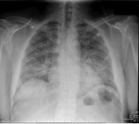
Figure 2
Diffuse bilateral infiltrates in a patient with pneumocystis jiroveci pneumonia (figure 2)
Treatment – General Considerations
Even though pneumocystis is classified as a fungus, it does not respond to antifungal treatment. treatment of choice is trimethoprim-sulfamethoxazole (cotrimoxazole). prednisone is added in severe pcp (pao2 <8kpa). Clindamycin plus primaquine is a 2nd line alternative, although primaquine is subject to section 21 approval and is rarely available. Although there are a few reports of successful caspofungin administration, based on the activity against the inclusion of (1 – 3) beta-d glucan into the fungal cell wall, clinical data are lacking and it is not a recognized treatment. Treatment should not be delayed if a pneumocystis pneumonia is suspected. After therapy is started the pneumocystis persist in the host for days to weeks. The treatment of the extrapulmonary manifestations of pneumocystis is the same as for the pneumonia. Patients without hiv usually show a response to treatment within 4 – 5 days and for hiv infected patients there should occur a response to treatment within the first 8 days. Considering antibiotic therapy, the recommended duration of treatment for hiv infected patients is 21 days and 14 days in all other patients. HIV infected patients tend to have a higher organism burden and could the response to treatment be slower, therefore a longer duration of therapy is required.
References
FIDSSA Members can earn CPD points by logging into the secure section of the website and visiting the MyCPD section.
Atlasville, Boksburg
South Africa
2022 © FIDSSA - All rights reserved • Website Terms of Use • Privacy Policy • Powered by E2
Admin login | Website login |
MYMEMBERSHIP®