
Moherndran Archary, UKZN. SASPID
An eight year old, HIV seronegative girl presented to an outpatients unit with a 3 month history of left sided neck swelling and intermittent fever. She had no TB contacts nor had been treated for previously TB. Her past medical history revealed 2 previous hospital admissions; the first, at 12 months of age was for an uncomplicated febrile convulsion, the second for a swelling on neck which was biopsied and treated with a short course of antibiotics. No further details were available and the swelling resolved completely within a few days, according to her parents.
On examination she was afebrile, there was significant unilateral cervical lymphadenopathy. She had multiple, non- matted, non- tender nodes in left anterior triangle of the neck, the largest of which was 1.5 x 1.5 cm. There was mass measuring 3 x 3 cm in the left submandibular region, which was mobile and non-tender, with a healed sinus.
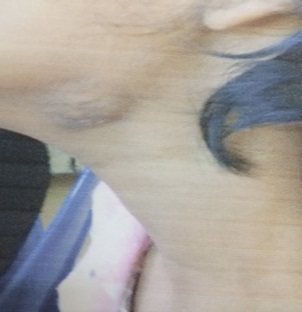
She had no lymph nodes palpable elsewhere. Her ENT examination was normal and she had no signs to suggest an upper respiratory tract infection. The respiratory, cardiac and abdominal system examination was normal.
Blood investigations, including an ESR were normal. Chest X-rays showed no radiological evidence to suggest Mycobacterium tuberculosis. The auramine stain was negative, but the TB culture from a fine needle aspirate of the submandibular mass was positive after 72 hours.
A PCR/Line probe Assay confirmed Mycobacterium fortuitum.
An excisional lymph node biopsy was done by the surgical team. The histology showed features of necrotising and non-necrotising granulomatous inflammation.
She was commenced on combination therapy of ciprofloxacin and clarithromycin for a minimum of 6 months duration.
On follow up she was well, with minimum scarring noted at the biopsy site.
Question: Discuss the aetiology of cervical lymphadenitis in chidren and the management of non-tuberculous mycobacterial infection.
Paediatric Non-Tuberculous Mycobacterium Cervical Lymphadenitis
Tuberculous and Non-Tuberculous Mycobacterium (NTM) are common causes of cervical lymphadenitis in the paediatric population.
Mycobacterium fortuitum is classified as a rapidly growing NTM. It is rarely isolated from the lymph nodes as compared to Mycobacterium avium complex (MAC), a slow growing NTM, which has been commonly isolated in the last few years.
NTM cervical lymphaditis distinguishes itself by presenting as a localised unilateral lymphadenitis. The anterior cervical and or submandibular lymph node complexes are more frequently affected.
Typical age of presentation is between 1-5 years. Although the reasoning remains unclear, it has been hypothesised that ‘children in this age group are more likely to ingest NTM organisms through oral exploration of objects that have been exposed to colonized soil or water.’
Patients are usually immunocompetent, with no constitutional symptoms. Some may experience mild fever with little or no tenderness over the area affected. The common presentation is a unilateral swelling in the neck. It can increase from 1cm up to 6cm in size, with central parts of the granulomatous lesions becoming necrotic and eventually fluctuant. The nodes can rupture and drain through a sinus tract in the skin.
The differential diagnosis of NTM cervical lymphadenitis includes Mycobacterium tuberculosis, pyogenic abscess, Cat Scratch Disease (Bartonella henselae), histoplasmosis, toxoplasmosis, infectious mononucleosis, salivary duct stones or tumours and lymphoma.
Blood investigations are usually unremarkable, a few patients may have a leucocytosis, the ESR may be elevated (although this is difficult to interpret in the setting of HIV due to high ESR being common in asymptomatic patients). A significant proportion of patients will have a non-reactive Mantoux test.
Given the prevalence Mycobacterium tuberculosis in our setting, a high incidence of suspicion should be maintained in any child presenting with cervical lymphadenitis. Mycobacterium tuberculosis should be excluded especially in children older than 12 years of age, if there is bilateral lymph node involvement with constitutional symptoms.
Fine needle aspirates (FNA) is considered an important diagnostic tool by some clinicians however the risk of chronic sinus formation and scarring post-FNA have made others less likely to perform this procedure.
Surgical treatment of choice is an excisional biopsy of the lymph node complex involved. This usually results in complete resolution of disease in a majority of the patients affected.
Histology of the excised lymph mode will reveal caseating or non-caseating granulomas. Acid-fast bacilli are observed in less than half of the specimens examined. Isolation of NTM from the lymph node or via molecular detection usually confirms the diagnosis.
A combination approach of both surgery and a course of antibiotics are practised by some clinicians. Antimicrobial susceptibility is usually indicated prior to commencement of treatment, since NTMs are resistant to first line agents used to treat Mycobacterium tuberculosis.
The inducible erm gene present in M. fortuitum can confer resistance to macrolides, hence macrolide monotherapy is usually discouraged. NTM lymphadenitis caused by MAC can be treated empirically with clarithromycin and rifabutin. Duration of treatment can vary with individual patients, extent of disease and response to treatment. 6 months is the standard recommended duration of treatment.
M. fortuitum infection in immunocompetent patients is usually associated with low mortality, however in immunocompromised patients it can result in deep organ involvement and disseminated disease.
FIDSSA Members can earn CPD points by logging into the secure section of the website and visiting the MyCPD section.
Atlasville, Boksburg
South Africa
2022 © FIDSSA - All rights reserved • Website Terms of Use • Privacy Policy • Powered by E2
Admin login | Website login |
MYMEMBERSHIP®