
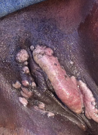
A 26-year old woman presented with a left swollen labia and massive genital warts around the vulva and labia. She was 6 months pregnant. No genital samples were taken as the patient declined.
Question 1: What is the epidemiology and clinical presentation of HPV infection?
Answer to Q1
Dr Richard Shope at The Rockefeller University, New York first considered a viral aetiology for genital warts in the 1930s. He noted that these viruses could infect human and cause papillomas (benign epithelial growths) such as skin and genital warts.
Genital human papillomavirus (HPV) infection is one of the most common sexually transmitted infections (STIs) worldwide, although maternal/fetal transmission may also infrequently occur.
Although the prevalence of HPV in women with normal cytological findings is high and variable across world regions, HPV-16, -18, -31, -52 and -58 are the most common types. Those classified as group 1 and carcinogenic to humans are HPV-16, -18, -31, -33, -35, -39, -45, -51, -52, -56, -58, -59, but HPV-16 account for >22% of HPV-positive infections. The estimate varies by geography and age with African regions showing higher average HPV prevalence ranging from 1.6% - 41.9%. The age distribution of cervical HPV infection peaks at younger ages, with lower prevalence in middle age due to changes in sexual behavior and becomes much higher at age >45 years and during peri-menopausal period due to higher rates of HPV persistence. The highest rates of genital infection are found in adults 18-28 years of age. It is estimated that 11.7% of women with normal cytological findings carry a detectable cervical HPV infection. The detection of HPV infection has been found to start consistently with a peak just after sexual debut, usually from 15 years of age.
HIV-infected women have a high prevalence of HPV infection and are infected with a broader range of HPV types than HIV-seronegative women. The proportion of HIV-infected women with HPV-16 has risen with increasing severity of cervical lesions. The immune suppression by HIV infection also appears to worsen the outcome of HPV infection and women infected with HIV are at significantly increased risk for invasive cervical cancer. Also, HPV infections are more likely to persist in HIV-infected women than in HIV-seronegative women and this persistence contributes to a higher prevalence of HPV infection among the HIV-infected women. It is also suggested that HIV-infected women without cytological abnormalities may be infected with a broader range of HPV types than HIV-seronegative women.
Although risk factors for infection are difficult to assess because of high frequency of subclinical infection, it is clear that major risk factors for acquiring genital HPV infection involve sexual behavior, particularly multiple sex partners. Other possible risk factors for acquisition of genital HPV infection include oral contraceptive use, pregnancy and impairment of cell-mediated immunity.
The spectrum of disease caused by HPV includes
Question 2: Discuss the differential diagnosis and investigations would you do to confirm the diagnosis?
Answer to Q2
The vast majority of anogenital warts are accurately diagnosed by clinical inspection. When combined with colposcopy, the acetowhite test (painting the genital skin or mucosa with 3% acetic acid to highlight flat condylomata) can help guide cervical biopsy.
The differential diagnosis of anogenital warts in sexually active persons, at risk of STIs include:
Biopsy and histology are occasionally required for diagnosis. Although visible anogenital lesions are present in some persons infected with HPV, the majority of individuals with HPV genital tract infection do not have clinically apparent disease. Conventional viral detection assays, including serologic assays and growth in cell culture are not available for the diagnosis and tracking of HPV infection.
Papanicolaou tests are a valuable screening tool, but they miss a large proportion of HPV-infected persons. Accordingly, HPV DNA detection assays have become a key research tool in the detection of HPV infection, particularly in asymptomatic individuals (Southern blot, PCR, Hybrid Capture). Analysis of cloned HPV DNA has allowed the exploration of the viral genome and the examination of the regions that may be important for carcinogenesis, accurate comparisons of different HPV types and an attempt at the formulation of HPV vaccine.
The inability to grow HPV in tissue culture results from HPV’s need for terminally differentiated epithelial cells (which no longer divide) in order to replicate. PCR or Hybrid Capture 2 for HPV detection in women with normal cytological findings is frequently used in most laboratories. There are also different sensitivities and specificities within PCR-based methods mainly due to differential sensitivity of PCR primers sets to specific HPV types with diverse prevalence of 32.1% to 76.0%. Validated HPV-PCR primers do not amplify all individual types in multiple-type infection. Certain types may be underestimated in some regions relative to others where more sensitive techniques were used. The type-specific performance of the assays depends not only on the technique but also on the laboratory and the processing of the specimen. Thus, cervical sampling techniques, cell transport medium, PCR DNA assays and HPV genotyping need to be standardized for diagnosis and is crucial for HPV vaccine surveillance and comparisons. Though cytology is important for screening and may reduce the burden of pre-cancerous lesions related to persistent HPV infections, it is subjective, poorly reproducible with limited sensitivity and repetitive for efficacy.
Question 3: How would you manage the case and what treatment modalities are there to manage HPV infections?
Answer to Q3
Many therapies are available to treat genital warts. Conventional therapies destroy wart tissue by cytotoxic or physical means. The Cytotoxic agents include:
while the physically ablative therapies mostly used are:
Although initially effective, the rate of recurrence is high due to the presence of the virus in adjacent normal-appearing tissue. Topical immune modifier Imiquimod in patients with external genital warts was once promising but has recently been shown to be ineffective in most cases. It acts by stimulating the immune system to produce high levels of interferon and other cytokines, which have antiviral, antiproliferative and immunomodulatory activities.
An integral component of therapy for patients with genital genital warts is patient counseling. Many patients respond to the appearance of genital warts with a strong mix of emotions, ranging from embarrassment to anger to fear. Genital warts can damage the patient’s feelings of self-esteem and interactions with sex partners. Worry about the possibility of developing cancer or of transmitting the disease to sex partners or to a newborn during vaginal delivery can also cause anguish.
Perhaps the most effective means for managing HPV disease would be a vaccine that prevents the occurrence of genital warts. The regional and country-based type-specific HPV prevalence may provide baseline values against which the global impact of HPV vaccination might be assessed in the future. A potential difficulty with vaccines for genital warts is that more than 20 different types of HPV can cause genital lesions. Accordingly, effective vaccination will require either the identification of a common antigenic epitope or the development of a multivalent vaccine directed against the relevant HPV types. Vaccine-targeted types 16 & 18 are the most frequent types worldwide and responsible for 70% of all invasive cervical cancers other oncogenic types are 52, 31, 58, 39, 56 and 31. A quadrivalent HPV-6/11/16/18 vaccine for the prevention of cervical, vulvar and vaginal intraepithelial lesions and genital warts contains some of these cancer-inducing strains. It was hypothesized that HPV-16 and 18 generated by vaccination may be able to bind to and possibly neutralize virions of HPV types closely related to 16 and/or 18, thereby preventing infection and disease associated with these other types i.e. giving cross-protection established by observed reduction in the incidence of cervical intraepithelial neoplasia (CIN).
References:
The patient responded well to our first communication and was referred for the commencement of ART. Subsequently the patient was lost to follow-up due to her reluctance to communicate with us further, thus no further follow-up of the patient was possible. Treatment with chemo-radiotherapy in this case would have resulted in complete resolution of the disease and due to pregnancy there may be a need for surgical intervention.
FIDSSA Members can earn CPD points by logging into the secure section of the website and visiting the MyCPD section.
Atlasville, Boksburg
South Africa
2022 © FIDSSA - All rights reserved • Website Terms of Use • Privacy Policy • Powered by E2
Admin login | Website login |
MYMEMBERSHIP®