ABOUT FIDSSA

Case of the Month

January 2012

A 40-year old immigrant from Democratic Republic of Congo presented to a Cape Town hospital following a seizure. He had flown to South Africa 1 year previously from his home in Kibibi village, Bandudu, DRC and had been well until 4 months prior to admission. His friend had noted a slowing of his thought processes, and increased somnolence.

One week prior to admission, he developed fever and shivering. Examination reportedly revealed a thin man with slowed affect, a temperature of 38.4oC and firm posterior cervical lymphadenopathy. Chest x-ray showed bilateral hilar adenopathy, but no pulmonary infiltrate. 4th generation HIV ELISA – negative.

Lumbar puncture:

Lymphocytes 49, polymorphs 0, red cells 0
Protein 0.77, glucose 4.2 (blood glucose 6.1)
Cryptococcal latex antigen test – negative
Acid fast bacilli - negative.
FTA and VDRL – negative

He was started on empiric therapy for TB meningitis with Rifafour (rifampicin, isoniazid, pyrazinamide and ethambutol) plus prednisone.

One month later, he was transferred to a tertiary level hospital, having failed to respond to therapy. On presentation, he was somnolent, his speech was markedly slowed and he was disorientated in time, place and person. He had a Parkinsonian tremor of the upper limbs, but no other focal neurological deficit. Posterior cervical chain lymphadenopathy was present and there was no change in his chest x-ray appearance. Ultrasound scan of the abdomen revealed retroperitoneal adenopathy and a bulky spleen measuring 12.5cm

Repeat lumbar puncture showed 20 lymphocytes, 14 polymorphs, protein 0.57 and glucose of 2.7.

Question 1: What is the diagnosis and construct a differential diagnosis for this man’s presentation?

Answer to Q1

This man’s presentation combined with his origin from a country endemic for human African trypanosomiasis (HAT, Figure 1) has all the hallmarks of stage II HAT secondary to infection with Trypanosoma brucei gambiense (T. b. gambiense)


Figure 1: Geographical distribution of West and East African trypanosomiasis


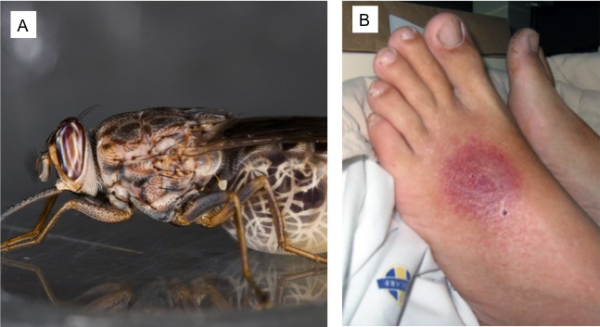
Unlike HAT secondary to Trypanosoma brucei rhodesiense (T. b. rhodesiense), which causes East African trypanosomiasis, T. b. gambiense is a more indolent infection with neurological manifestations indicative of CNS penetration by the typanosome occurring months to years after initial infection. HAT is transmitted by tsetse flies (Glossina spp, Figure 2A), and at the site of infection, a trypanosomal chancre (Figure 2B) may form after about 5 days, although this is less common in West African trypanosomiasis than in East African Trypanosomiasis. Thereafter, stage I disease follows ~2-3 weeks later in West African disease, characterized by haematolymphatic dissemination of trypanosomes, with clinical features of fever, non-specific symptoms, transient rashes and lymphadenopathy (including posterior cervical chain lymphadenopathy, first described by Winterbottom and known as Winterbottom’s sign). Hepatosplenomegaly may also be evident. Symptoms may vary in cycles of one to several days as a result of the parasite changing its variant surface glycoproteins.


Figure 2: Tsetse fly (A) and (B), a trypanosomal chancre in a patient with East African trypanosomiasis (courtesy of Prof Lucille

If untreated, stage II disease occurs with neurological involvement months to years later, although this may occur within weeks in T. b. rhodesiense infection, and presents in a far more acute manner. The clinical features of 500 Angolan patients recruited into a randomised melarsoprol dosing study with stage II T. b. gambiense infection are shown in Table 1. Early targeting of the thalamus, choroid plexus, pineal and hypophyseal regions by trypanosomes explain some of the initial neurological features. Our patient manifested Parkinsonian features indicative of basal ganglia involvement, somnolence and impaired speech. Although the total sleeping duration remains largely unchanged, the pattern changes, with diurnal somnolence, nocturnal restlessness and insomnia common. Periods of sleep are generally shorter and spread out over the 24 hours.


Table 1: Neurological manifestations of T. b. gambiense infection

Differential Diagnosis:

The differential diagnosis of stage II T . b. gambiense infection in the setting of sub-Saharan Africa includes many of the causes of chronic meningoencephalitis. Diagnoses that should be considered include:

  1. Infections
    1. Syphilitic meningomyelitis
    2. Cerebral tuberculosis
    3. HIV-associated infections
      1. HIV-associated neurocognitive disorder (HAND)
      2. HIV encephalitis
      3. Progressive multifocal leukoencephalopathy
      4. Cryptococcal meningitis
      5. Lymphoma
    4. Chronic viral encephalitis
  2. Non-HIV-associated malignancy
  3. Metabolic encephalopathies
  4. Psychiatric illness

Cerebral malaria and bacterial meningitis are unlikely differential diagnoses of T. b. gambiense stage II disease, although their acute presentation would form part of the differential diagnosis for T. b. rhodesiense stage II disease.

Question 2: How would you diagnose and treat this infection?

Answer to Q2

Diagnosis of T. b. gambiense stage II HAT may be by parasitological and/or serological means. The most common test used in screening is the card agglutination test for trypanosomiasis (CATT, Figure 3), which uses reagent containing stained freeze-dried trypanosomes and has high sensitivity and specificity.


Figure 3. Card agglutination test for trypanosomiasis (CATT)

Parasitological diagnosis involves examination of chancre or lymph node aspirate, CSF and blood for the presence of trypanosomes. Concentration methods increase the chance of detection, as parasite numbers in body fluids in T. b. gambiense infection are very low.

Lymph node or chancre aspirate – wet prep examination of fluid examined at x400

Blood – thick and thin films (Figure 4A), microhaematocrit centrifugation based on examination of the buffy coat zone, quantitative buffy coat (QBC) technique using a glass haematocrit tube precoated with acridine orange and anticoagulant, and miniature anion-exchange centrifugation technique (m-AECT) have all been employed

CSF – centrifugation and microscopy. CSF examination should include a search for the presence of morula cells (cells of Mott, figure 4B), which are abnormal plasma cells containing rough endoplasmic reticulum-derived vesicles containing IgM immunoglobulin. Morula cells are not pathognomonic of HAT, but also occur in multiple myeloma, autoimmune disorders and we have observed them in a patient with HIV (unpublished observation)


Figure 4. T. b. gambiense in blood (A) and a morula cell (B) in the CSF

Treatment of HAT

Drugs used to treat the first stage of HAT are trypanocidal but do not cross the blood brain barrier (BBB), whereas drugs used to treat stage II disease need to cross the BBB. Table 2 shows the drugs commonly used to treat HAT


Table 2: Treatment of HAT (second line drugs are depicted in brackets)

*Pentamidine has some activity against EAT and may be used if suramin is unavailable

Until the development of eflornithine, the mainstay of treatment for stage II T. b. gambiense infection was melarsoprol, an arsenical derivative that causes reactive encephalopathy in 5-10% of treated patients and is fatal in 10-70% patients who develop this complication. Eflornithine is efficacious and better tolerated than melarsoprol. Main side effects include seizures, gastrointestinal disturbance and bone marrow suppression, all of which are reversible on discontinuation of the drug.

A recent non-inferiority RCT of stage II patients in the DRC showed that combination therapy of nifurtimox and eflornithine (NECT) for 10 and 7 days respectively was non-inferior to standard, longer duration eflornithine monotherapy. The simpler, shorter regimen, coupled with the advantage of reducing the risk of development of resistant parasites makes NECT a useful alternative to longer course monotherapy.

Outcomes and Lessons Learnt

Stage II T. b. gambiense infection was confirmed on examination of a buffy coat sample, which showed a single trypomastigote (Figure 5A). Neither lymph node aspirate nor CSF showed evidence of trypomastigotes. Morula cells were abundant in his CSF (Figure 5B), which showed a raised IgM level.


Figure 5: Trypanomastigote in buffy coat (A) and morula cells in CSF (B)

The card agglutination test for trypanosomiasis (CATT) confirmed disease caused by T. brucei gambiense. Eflornithine was procured through the World Health Organization Programme for Neglected Tropical Diseases, Geneva and he was treated with 100mg/kg ivi 6 hourly for 14 days. He made an excellent clinical recovery, being fully conversant, mobile and without tremor on discharge.

Southern Africa has a large number of immigrants from trypanosomiasis-endemic countries. Stage II West African trypanosomiasis should be considered in migrants and travellers from endemic countries in the differential diagnosis of the patient presenting with chronic meningoencephalitis.

Earn CPD Points

CPD QUESTIONS

FIDSSA Members can earn CPD points by logging into the secure section of the website and visiting the MyCPD section.