
Dr Rena Hoffmann, Microbiologist, National Health Laboratory Service, Tygerberg Hospital
A 44-year old man was referred to Tygerberg Hospital with one-month progressive right-sided weakness, speech impairment and new onset convulsions. His medical history is remarkable for longstanding alcohol dependence with poor social circumstances. Clinical evaluation at the time of admission confirmed a right-sided hemiplegia with aphasia. He remained afebrile during his hospital stay and intermittent right-sided focal convulsions were observed in the ward.
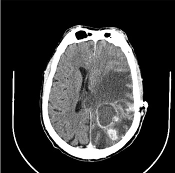
On initial special investigations, the white blood cell count was within normal limits and HIV serology was negative. A contrasted CT-scan of the brain demonstrated a left parietal brain abscess resulting in midline shift.
An emergency craniotomy was performed and 40ml of non-offensive pus was drained. Based on the findings at craniotomy, empiric ceftriaxone, metronidazole and Rifafour were started. His focal seizures were controlled with phenytoin.
Pus was sent for routine Microscopy, Culture & Sensitivity (MC&S) as well as Mycobacterial MC&S. The Gram stain showed a moderate amount of neutrophils, but no bacteria were observed. Two days later the aerobic bacterial culture yielded an organism with features suggestive of a Nocardia species. The Gram stain of these colonies showed delicate Gram-positive filamentous branching bacilli. A modified Ziel Neelsen stain of the isolate was positive. The colony morphology on the blood agar plate was chalky white and had the aroma of wet soil, which although not pathognomonic, is suggestive of Nocardia). Based on these findings and current Western Cape Academic hospitals antimicrobial recommendations, high dose Trimethoprim-Sulphamethoxazole (TMP-SMX), one tablet (80/400mg) for each 4 kg body weight per day was started. Subsequent PCR and DNA sequencing of the 16SrRNA gene confirmed a Nocardia abscessus infection. Antimicrobial susceptibility testing (E-test method; AB Biodisk Solna, Sweden) confirmed that the isolate was sensitive to TMP-SMX with a MIC of 0.03µg/ml. The ceftriaxone, metronidazole and Rifafour were discontinued.
The patient’s neurological deficits resolved after 6 weeks of treatment and he was discharged on a lower dose of TMP-SMX and is being followed up on an outpatient basis.
Question 1: What are the main clinical manifestations of infection with Nocardia species?
Answer to Q1
Question 2: What are the risk factors for developing disease caused by Nocardia?
Answer to Q2
Nocardiosis is typically regarded as an opportunistic infection, but approximately one-third of patients do not have a recognized immunodeficiency. Cell-mediated immunodeficiencies are usually associated with nocardia infections, the most common being glucocorticoid therapy, malignancy (solid tumors and hematological malignancies), organ and hematopoietic stem cell transplantation, and advanced HIV infection. Other conditions that have been associated with nocardiosis include diabetes mellitus, alcoholism, chronic granulomatous diseases, alveolar proteinosis, tumor necrosis factor-alpha inhibitor therapy, chronic obstructive pulmonary disease and tuberculosis.
Question 3: How does CNS Nocardia infection present?
Answer to Q3
Clinical manifestations of CNS nocardiosis usually result from local effects of granulomas or abscesses in the brain, and less commonly, the spinal cord or meninges. Disease frequently progresses over months to years and causes a broad range of neurological deficits, including chronic behavioral and psychiatric disturbance, which reflect localization in the cerebral cortices, basal ganglia and midbrain.
Question 4: What is the differential diagnosis of CNS Nocardia infection?
Answer to Q4
Question 5: What are the treatment modalities for Nocardia brain abscesses and what is the recommended duration of therapy?
Answer to Q5
Successful therapy requires the use of antimicrobial drugs and, in some cases, appropriate surgical drainage.
Surgery should be performed when abscesses are accessible and relatively large, the patient’s condition deteriorates or lesions progress within 2 weeks of therapy, or there is no reduction in abscess size within 1 month. Decompression of lesions can be accomplished by stereotactic aspiration, although cure in many cases is affected only after craniotomy and total excision. Small abscesses can be cured by prolonged antimicrobial therapy. As abscesses may progress in the face of appropriate therapy, all patients must be monitored frequently with neuro-imaging.
Sulfonamides are still considered to be the mainstay of therapy for Nocardia infections and most clinicians prefer TMP-SMX. Combination therapy is recommended, particularly in severely ill patients, those with cerebral involvement or disseminated nocardiosis, and immunosuppressed patients. Empiric combination therapy should be commenced pending susceptibility test results. Three-drug regimens consisting of a sulfonamide, amikacin, and either a carbapenem or third-generation cephalosporin are suitable for empiric therapy for the treatment of high-risk patients.
Susceptibility testing is especially indicated when patients present with deep-seated or disseminated infection, fail to respond to initial therapy or relapse after therapy, and when alternatives to sulfonamides are being considered. Testing is also indicated when relatively resistant Nocardia species, such as N. farcinica, or one of the newly described species, such as N. abscessus, has been identified.
Alternative therapies for Nocardia infection in general include the combination of amikacin and TMP-SMX, imipenem and amikacin, meropenem, ceftriaxone, cefotaxime and cefuroxime, minocycline, linezolid, moxifloxacin and tigecycline. Ertapenem is inactive against Nocardia.
HIV-seronegative and other immunosuppressed patients should be treated for 6 to 12 months, depending on the underlying state of immunosuppression and response to therapy. Treatment should be continued for 12 months or longer if there are intercurrent increases in immunosuppression. For patients who must be maintained on steroid or cytotoxic therapy, prolonged low-dose maintenance therapy may be required.
In HIV-infected patients, early institution of a prolonged primary course of anti-nocardia therapy is essential. Patients present late presentation and relapse may occur. Secondary prophylaxis should be continued for at least 6 months after the CD4 count rises above 200 cells/mm3. Some experts recommend life-long secondary prophylaxis.
References
Nocardia infection should be considered in the differential diagnosis of patients with impaired cell-mediated immunity who present with clinico-radiological evidence of brain abscess. Species identification and antimicrobial susceptibility are recommended in order to identify more resistant species and to confirm TMP-SMX susceptibility.
FIDSSA Members can earn CPD points by logging into the secure section of the website and visiting the MyCPD section.
Atlasville, Boksburg
South Africa
2022 © FIDSSA - All rights reserved • Website Terms of Use • Privacy Policy • Powered by E2
Admin login | Website login |
MYMEMBERSHIP®