
Frans Radebe, NICD
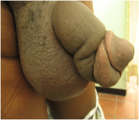
A 49-year old man presented to Alexandra Men’s clinic, Gauteng on the 13/08/2011 with a swollen penis of one month duration, associated, swollen, enlarged testes and swollen legs. Originally from Tzaneen, Limpopo, he lives in Johannesburg, visiting home on a monthly basis. He had not travelled outside of South Africa. His HIV status was unknown (he refused testing) and he was unwilling to give a sexual history, other than the fact that his last sexual encounter was 1 month previous with a single partner. He denied trauma to the penis, genital ulceration or penile discharge. On examination, he manifested a ‘Saxophone- like penis” (figure 1), which was cold, non-tender, enlarged and curved. There was no urethral discharge, penile or perianal ulceration, or regional lymphadenopathy. The scrotum was huge and swollen and testes were unpalpable. He had bilateral pitting oedema of the legs and his blood pressure was 157/94 mmHg. The rest of the examination was unremarkable.
The attending physician ruled out sexually transmitted infections (STIs) such as syphilis, lyphogranuloma venereum (LGV) and donovanosis on the basis of there being no clinical evidence of genital ulceration or urethral discharge. The patient was referred to a urologist for further assessment and treatment.
Figure 1 Genital elephantiasis resembling a “saxophone penis”
Question 1: What is the differential diagnosis of “Saxophone” penis?
Answer to Q1
Genital elephantiasis due to STIs
Non-STI causes of genital elephantiasis:
Question 2: Discuss the pathogenesis of STI-related and non-STI-related genital elephantiasis.
Answer to Q2
STI-related genital elephantiasis
Genital elephantiasis due to STIs is most commonly seen as part of the tertiary stage of Lymphogranuloma venereum (LGV) due to Chlamydia trachomatis serovars L1-3. It is more common among high-risk HIV-infected men who have sex with men (MSM)1, due to sero-sorting and an increased probability of transmission due to reduced condom use and high risk sexual practices. LGV classically affects the lymphatics, starting as a small, transient, primary lesion(s) followed by development of suppurative regional lymphadenitis. Progression to the tertiary stage occurs in about one quarter of patients, typically in women and MSM. There is potentially, a long latent period between primary infection and tertiary manifestations such as Saxophone penis, ranging from 1-20 years2. Saxophone penis results from progressive lymphangitis giving rise to indurated oedema and sclerosis of underlying subcutaneous tissues. It is hypothesized that there is differential contraction of connective tissue on the dorsal and ventral aspects of the penis determined by differential blood supply, leading to bending of the penis dorsally2. The corpora cavernosa attaches to the ischial tuberosity anchoring it, whereas the bulbospongiosis and glans penis that lack such an anchor are bent upwards. The dorsal curvature is further enhanced by differential extravasation and oedema of the ventral prepuce.
Donovanosis is the second commonest STI-related cause of Saxophone penis and genital elephantiasis in general. Unlike the thrombolymphangitis and peri-lymphangitis that initiate the pathology associated with LGV-induced genital elephantiasis, Klebsiella granulomatis infection leads to lymphatic constriction, trapping the chronic granulomatous inflammatory response and causing the penis to twist along its long axis3.
Penoscrotal elephantiasis due to syphilis is well described but very rare. One case reported oedema and penile deformity one month after unprotected intercourse and at the time, a chancre was still present with atypical rash. The likely pathogenesis of the oedema is due to lymphatic obstruction due to enlarged inflamed lymph nodes4.
Non-STI-related genital elephantiasis
Genital elephantiasis due to non-STI-related causes is most commonly associated with endemic lymphatic filariasis, caused in 90% of cases by Wuchereria bancrofti with Brugia Malayi responsible for the majority of the remaining cases. Infection results from transmission of infective larvae from culex or anopheline mosquitos, gaining access to the peripheral lymphatics and moving centrally to the lymphatics proximal to the draining lymph node. Following a moult, the juvenile filariae become adult worms, pairing and mating. Adult worms live in ‘nests’ with females producing microfilariae, which enter blood and lymph, to be taken up by mosquitos.
Chronic lymphatic filariasis is a late stage of the infection seen mainly in endemic populations exposed to repeated infection. These repeated attacks during which lymph nodes become inflamed are followed by severe retrograde painful lymphngitis. Initially, acute oedema resolves, but eventually, chronic lymphatic obstruction ensues. The end-stage manifestation of lymphoedema and elephantiasis most commonly affects the legs, scrotum, and penis. Lymphatic filiariasis that is transmitted by Aedes spp is commonly associated with upper limb and breast oedema due to the favoured distribution of Aedes biting these areas. Treatment of lymphatic filariasis is with albendazole and ivermectin in onchocerciasis-endemic areas or albendazole and DEC in non-onchocerciasis-endemic areas. Scrupulous hygiene is recommended to prevent secondary infection.
Primary lymphedemas are an uncommon cause of genital elephantiasis, are predominantly found in females, are congenital or familial and usually appear at puberty or after 35 years of age5. It is a disabling condition and emotionally incapacitating. Lymphedema of the male genitalia is caused by reduced lymphatic flow, with subsequent enlargement of the penis and scrotum. Swelling is characterized by severe discomfort and a progressive loss of sexual and urinary function, which cause more emotional stress making surgical intervention imperative6.
Question 3: Discuss the management of a patient manifesting Saxophone penis.
Answer to Q3
Optimal management of a patient manifesting Saxophone penis or any type of genital elephantiasis would begin with a thorough history including sexual and travel history. Family history and any prior episodes of genital oedema should be sought. Constitutional symptoms may be present, particularly in rare cases due to genital tuberculosis. Laboratory investigation may include any of the following tests depending on the history and suspected aetiology:
An interdisciplinary approach to treatment is required involving physicians, urologists and a dermatologist opinion may be needed.
The main objectives of treatment are to reduce swelling, restore shape and normal sex function, and prevent inflammatory episodes2,3.
Although the role of antibiotic therapy to treat LGV-related penoscrotal lymphoedema is questionable, most experts would give a 14-day course of doxycycline empirically especially if LGV serology is positive. Azithromycin is the antibiotic of choice if donovanosis is confirmed.
Physical compression therapy methods are challenging, rarely produces a lasting result. Most patients will require a urological +/- plastic surgical consult. Surgery (bypass or reduction procedures) under the cover of appropriate antibiotics for those patients in whom the disorder is disabling and persistent3,6 may involve extensive resection of the involved tissue, scrotoplasty and total excision of lymphedema tissue of the penile shaft.
References
Other Reading:
The patient was referred to Edenvale General Hospital, Gauteng for further assessment and treatment, the details of which are unavailable. Telephonic follow-up on 22/08/2011, indicated that the testes and penis were still swollen, but had improved, and his bilateral leg oedema was still present. As we couldn’t establish the treatment regimen given to the patient, he was requested to visit the STI clinic again for further investigation and follow-up.
Saxophone penis is one manifestation of genital lymphoedema or ‘elephantiasis’. It may be a sequel to late stage STIs, most commonly LGV, or in endemic areas, may result from lymphatic filariasis. Optimal management of patients presenting with a Saxophone penis will involve physicians with knowledge of STIs, urologists, plastic surgeons and psychologists as the psychological affects of such a deformity are often profound. This patient seems as yet, not to have been optimally screened or treated for STIs, however the role of antibiotics at this stage of the disease is questionable.
FIDSSA Members can earn CPD points by logging into the secure section of the website and visiting the MyCPD section.
Atlasville, Boksburg
South Africa
2022 © FIDSSA - All rights reserved • Website Terms of Use • Privacy Policy • Powered by E2
Admin login | Website login |
MYMEMBERSHIP®