
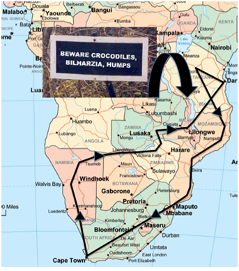
A 19 yr old, male, smoking, University student travelled overland from Cape Town to northern Namibia, through the Caprivi Strip to Victoria Falls and Lake Kariba, on through Zambia to Lake Malawi where he swam in Cape Maclear, Monkey bay and Kata bay, before continuing on to Dar es Salaam, Lake Tanganika and Gombe National Park to see the Chimpanzees. He returned to Cape Town via the Quirimbas archipelago and Musina da Praia in Mozambique. During his travels, which lasted 5 months, he and his girlfriend camped most of the time and occasionally, stayed in small guesthouses. They ate food from the roadside and took doxycycline chemoprophylaxis, although his adherence was poor (once every 3 days). He received multiple mosquito bites, but was unaware of any Tsetse fly bites or other insect exposures. Pre-travel vaccinations included Yellow Fever alone.
Two weeks after experiencing itching skin following a swim in Cape Maclear, he awoke with fever and chills associated with profuse watery diarrhoea 5-6 x /day and severe colic. Blood tests for malaria were negative, but he took Co-artem, under-dosing himself by half for the last 3 doses. Symptoms continued, but the diarrhoea resolved after 4 days, leaving him lethargic, febrile and and with a developing dry cough. Stool, blood and urine tests were negative. He was given a course of double-dose doxycycline, ciprofloxacin and azithromycin resulting in resolution of fever, but not the cough, which persisted and became productive of white sputum after 10 days. Sputum production cessed when he stopped smoking. 3 weeks after his first fever, he experienced headache, meningism, fever and worsening of his cough. He was diagnosed with typhoid fever on clinical grounds alone, receiving a 10 day course of ciprofloxacin and another course of Co-artem for good measure. Thereafter, there was a slow resolution of symptoms, although he was left with lethargy and a dry cough. He returned to Cape Town to seek medical advice.
At the time of presentation, his cough had resolved and he had started smoking and drinking again. Examination was non-contributory. He was afebrile, his chest was clear with no organomegaly.
Investigation revealed a total WBC 10.4 x 10⁹/L, but eosinophils were raised at 2.20 x 10⁹/L [normal 0.00-0.40]. The rest of his FBC was normal as was hepatic and renal function. CRP 11.6 mg/L. Chest X-ray was normal.
A clinical diagnosis of resolving acute schistosomiasis was made. Three filtered urine and 3 concentrated stool specimens were negative for schistosoma ova, but schistosoma IgG ELISA was positive, as was the IgM and IgA responses to cercarial antigens.
As his symptoms had resolved, he was given praziquantel 40mg/kg in 2 divided doses. Within hours of his second dose, his cough and fever returned with profound malaise. He was given prednisone 0.5mg/kg with an excellent symptomatic response within the 4 hours of the first dose. He was treated for 3 days in total with no relapse.
Question 1: How is the immunopathogenesis of schistosomiasis linked to the clinical presentation of disease and how does it impact on choice of treatment for acute schistosomiasis?
Answer to Q1
Schistosomiasis is caused by one of 5 schistosomes that infect humans; ‘Intestinal’ schistosomes S.mansoni, S. japonicum, S. mekongi and S. intercalatum, and the ‘Urinary’ schistosome, S. haematobium. Adult schistosomes of S. haematobium reside in the vesical and pelvic plexi of the inferior vena caval system, laying eggs (figure), which are either trapped in the bladder or genitourinary tissues, or expelled in the urine. In contrast, adult intestinal schistosomes reside in the peri-colonic venous plexi of the portal system and expel ova into the stool. On hitting water of 10-30C, the developing miracidium within the egg hatches and infects the snail host specific to that schistosome species. Asexual reproduction occurs within the snail, producing thousands of single-sexed cercariae (figure), the sex of which depends on the infecting miracidium. Cercariae are released from the snail and survive in water for 36-48h.
Following immersion in a cercarial-infested fresh-water environment (salt water does not support the intermediate snail hosts), humans can become infected by penetration of the cercaria through the epidermis and into the dermis. This causes a cercarial dermatitis, which in symptomatic persons, presents with a transient papular, itchy eruption (figure) that usually lasts a short period of up to 4-8 hours. Each papule corresponds to the reaction around penetrating cercariae, and patients may be infected by many hundreds during a single exposure.
Once the cercaria penetrates the epidermis, it looses its tail to become a schistosomule (Figure), the juvenile form of schistosoma. The schistosomule must traverse the basement membrane, enter the dermis and penetrate through the capillary walls into the bloodstream, which may take around 10-48 hours. Once in the circulation, the schistosomules reach the right side of the heart within 5-7 days, enter the pulmonary circulation, back to the left side of the heart and then into the systemic circulation. Some will find their way to the splanchnic capillaries, whilst others will re-circulate in the systemic circulation for an undetermined time. Those that traverse the splanchnic capillaries enter the hepatic portal system, where they feed on blood, increase in size and move upstream to larger vessels where they shorten and mature into adults. Adult pairing occurs ~28-35 days after infection. The female lies in the gynaecophoric canal of the male (figure) and paired adults migrate to their final resting places and copulate daily producing eggs that are excreted, trapped or embolize. Those eggs that are trapped in tissues elicit an granulomatous inflammatory response that causes distortion of normal tissue structure, fibrosis and polyp formation (intestinal schistosomiasis). Chronic urinary schistosomiasis may be silent, or present with haematuria, haematospermia, ureteric strictures ± hydronephrosis and as a late complication, is a risk factor for development of squamous cell carcinoma of the bladder. Chronic intestinal schistosomiasis may again be asymptomatic or present with bloody diarrhea and polyp formation. Granulomatous reaction and fibrosis around the peri-portal tracts leads to Symmers’ fibrosis, which may lead to an increase in portal pressure with variceal bleeding. Hepatic synthetic function is maintained, differentiating the portal hypertension from those that cause a true cirrhosis.
There has been a long-standing debate as to the exact cause of the clinical manifestations of acute schistosomiasis (AcS), between those that believe it results from an immune response to the start of egg laying (oviposition) by the adult female, versus a reaction to juvenile schistosomule antigens. AcS (Katayama Fever, Katayama Syndrome) most commonly presents in travellers, as fever, malaise, urticaria, respiratory symptoms including wheeze and eosinophilia. The terms Katayama Fever or Syndrome are no longer used as AcS can occur in infections other than those caused by S. japonicum, which was first described in Katayama, Japan, and secondly because fever is not a universal finding. Indeed, in some studies, only just over 50% of patients exhibited fever. Single exposure outbreaks in travellers have identified onset and duration of symptoms. Rash, itch and fatigue generally occur around 3 weeks after exposure, the latter being the longest-lived symptom. Cough and other respiratory symptoms may lag behind in onset, occurring up to 1-2 weeks later and continuing after the rash and fever have disappeared (as in our case). It is less well recognised in the endemic population and there is some evidence to suggest that maternal schistosomal antibodies might alter the immune response to first infection with schistosomal cercariae.
Circulating immune complexes are present in blood during AcS, but decline in chronic disease. Many of the features of AcS mimic serum sickness, which is mediated by soluble circulating immune complexes. Toxic eosinophil proteins released in lung, myocardium and CNS are also proposed to play a role in pathogenesis of AcS, as does the heightened pro-inflammatory mileu including raised TNF-a, IL-1 and IL-6 that predominates during early infection, before the increase in Th2 profile once egg-laying begins.
A recent analysis of single exposure outbreaks in groups of travellers1 shows that a substantial proportion of AcS occurs early after infection during the pre-patent period i.e. prior development of the adult worms and oviposition. This provides strong evidence in favour of the primary immune response being directed against circulating juvenile schistosoma antigens. Although praziquantel has shown limited activity against very early (week 1) juvenile schistosomules in murine models of infection, it is not active against later schistosomules, but rather, against adult schistosomes. Hence, it is not the treatment of choice for those presenting with AcS, the symptoms of which are driven by the reaction to schistosomule antigens. Rather, it is corticosteroids that treat the symptoms of AcS. There is no good evidence to dictate the dose of steroid that should be used, yet prednisone 0.5mg/kg for 3-7 days is often used to good effect. Some patients require longer courses.
Question 2: Why did the patient’s symptoms worsen with praziquantel?
Answer to Q2
There have been a number of reports of life-threatening exacerbation of symptoms in patients with AcS, treated with praziquantel2,3. These include cardiac and neurological manifestations, the latter seemingly driven by a vasculitic process. The exact cause of the deterioration that occurs in AcS patients treated with PZQ is undetermined. One hypothesis suggests that as the drug is active against very early juvenile schistosomules in murine models of S. mansoni infection, and seeing as though juvenile schistosomules can re-circulate for a prolonged period, re-circulating immature schistosomules may be susceptible or partially susceptible to the praziquantel. The exposure of tegument antigens that results from praziquantel treatment may then exacerbate the immune response. Praziquantel administration in patients presenting with features of AcS should be delayed until 3 months following last water exposure, by which time all remaining worms should have matured to adulthood and thus be sensitive to the drug. This should also reduce the chance of exacerbation of the clinical presentation.
Question 3: Are there any other drugs that could be used for patients presenting with acute schistosomiasis?
Answer to Q3
The most promising candidates in murine models of infection that are active against juvenile schistosomes are the artemesinin derivatives and mefloquine. Artemesinins have been used successfully in China to prophylax against S. japonicum infection during floods4. Subcutaneous infection of mice with cercaria, followed by mefloquine at either day 7,14,21,28,35,42 or 49 showed >80% reduction in worm burden even as early as when administered 7 days post-infection5. In contrast praziquantel reduced the worm burden by < 10% unless given from 35 days post-infection onwards and only reached a reduction of > 80% when given at day 49. This supports the understanding that praziquantel is active against mature adult worms rather than juvenile forms. Oxamniquine, a drug active against S. mansoni alone and used in South America, has also shown good activity in murine models6.
Both artemesinins (artesunate and arthemether) in combination with a variety of second agents, including mefloquine, have been trialled in the treatment of chronic schistosomiasis, reviewed by Utzinger et al in Lancet Infectious Diseases7. Variable cure rates and egg reduction rates have been reported.
Question 4: How should patients with acute schistosomiasis be followed up?
Answer to Q4
The options for follow-up of patients who present with AcS are limited by the fact that egg detection in urine or stool is often lacking either because oviposition has not yet begun, or as egg-laying may be intermittent, eggs are often not picked up in body fluids. Rectal snips to look for eggs may increase yield, yet it is an uncomfortable procedure and not available at many point-of-care sites. If eggs are present in urine or stool, then follow-up should include re-examination for eggs a month after praziquantel treatment. In addition, a reduction in eosinophil counts should be looked for, although persistently raised eosinophil counts have been documented even up to 1 year after praziquantel therapy. Once raised, antibody titers are not a reliable method of monitoring therapeutic response. A recent study in Australian travellers and immigrants showed that a 4-fold rise occurred in 28% of immigrants within the 1st year after treatment and a 2-fold rise occurred in 14% of travellers8. Only two thirds of travellers and one third of immigrants achieved a 4-fold reduction in antibody titres at 30 months post-treatment.
References
Although thankfully not life-threatening, this case exemplifies the clinical deterioration that can occur when treating patients presenting with AcS with praziquantel. Other case reports that have been published include life-threatening neurological and cardiac deterioration. Prednisone 0.5mg/kg should be the treatment of choice for patients presenting with severe manifestations of AcS, and praziquantel administration should be delayed until at least 3 months after the last risk-exposure.
FIDSSA Members can earn CPD points by logging into the secure section of the website and visiting the MyCPD section.
Atlasville, Boksburg
South Africa
2022 © FIDSSA - All rights reserved • Website Terms of Use • Privacy Policy • Powered by E2
Admin login | Website login |
MYMEMBERSHIP®