
Dr Philip Botha, Infectious Diseases Specialist, Tygerberg Hospital
facial flushing, headache, generalized myalgia and arthralgia, one day after arriving in South Africa on holiday. A blood smear for malaria parasites and a malaria antigen test were negative, but no additional investigations were requested. A presumptive diagnosis of Influenza was made and symptomatic treatment was prescribed. The patient developed a generalized rash and melaena stools 4 days later and was referred to the Emergency Unit of a Cape Town Hospital.
Apart from Thalassemia trait, there was no prior medical history. She lived in Bangkok, Thailand where she was employed as a teacher. She travelled to Shanghai for the purpose of work in the three months prior to visiting South Africa, but had returned to Bangkok for the week prior to travelling to South Africa. She received a Yellow Fever vaccination a number of years previously but did not receive other vaccinations recently and was on no regular medication. She had experienced multiple mosquito bites in Bangkok, but did not recall any other insect exposure, nor had she had any contact with animals. She had no risk factors for HIV infection and no contact with any person with fever or bleeding.
At the time of her presentation she was afebrile and haemodynamically stable. A diffuse erythematous macular rash with islands of sparing was noted (figure 1a&b). A tourniquet test was positive. There was no eschar, lymphadenopathy or hepatosplenomegaly. Evaluation of the cardiac, respiratory and neurological systems was non-contributory.
Fig 1a: Erythematous macular rash with island of sparing
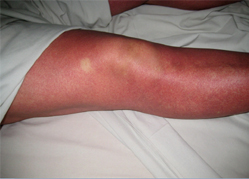
Fig 1a: Erythematous macular rash with island of sparing
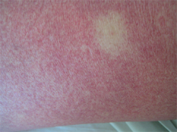
Fig 1b: Close-up image of island of sparing on the background of a diffuse rash
Special investigations:
White cell count 2.4 x 10⁹/L; haemoglobin 12.4 g/dL; platelet count 20 x 109/L.
Liver function tests: ALT 63 IU/L; AST 174 IU/L.
INR & PTT normal.
CRP 6 mg/L.
Blood & urine cultures negative.
Chest radiograph within normal limits.
Blood sample sent to the NICD in Johannesburg for further testing.
A clinical diagnosis of severe dengue with haemorrhage was made.
Question 1: What is the differential diagnosis?
Answer to Q1
The differential diagnosis of the traveller presenting with fever, rash and bleeding is extensive and includes both infectious and non-infectious causes. The initial critical step in the assessment is the patient’s epidemiological exposure to formulate a plausible differential diagnosis, exclude certain conditions and guide investigation. Combining the epidemiological exposure with results of basic investigations (full blood count, liver function tests and coagulation studies) will help to further narrow down the possible causes.
Since this patient flew directly from Thailand to South Africa, it is most likely that she contracted her illness in Bangkok, although a recrudescence of an infection acquired in the past is not ruled out. The overall picture, with exposure to mosquitoes in Thailand, her clinical presentation with a typical rash including ‘islands of sparing’ i.e. white patches surrounded by the macular or maculopapular rash, bleeding, leucopaenia and thrombocytopenia is most suggestive of an arbovirus infection such as dengue, the leading cause of fever in travellers outside Africa [1]. Chikungunya, another arbovirus currently circulating in Asia and Africa is often difficult to distinguish from dengue and is high on the differential diagnosis list. The likelyhood of her presentation being due to another viral exanthem such as measles or rubella will depend on her vaccination status, history of childhood infection and any close contact with a confirmed / suspected case which she did not have. Moreover, haemorrhage due to these infections would be uncommon.
Malaria is an important cause of fever in the traveller from Southeast Asia and patients should be aggressively investigated for this as untreated falciparum malaria may be rapidly fatal. A single negative blood smear and/or antigen test (as in this particular patient) does not rule out malaria, yet the rash was not consistent with DIC complicating severe malaria, and both Bangkok and Shanghai are considered malaria-free areas [2].
She was not sexually active and had no history of intravenous drug abuse which would increase her risk of acute HIV infection as a cause for illness. She did not own pets, but could have been in contact with excreta from rodents making leptospirosis part of the differential. Leucopaenia would be atypical in leptospirosis, as would a normal CRP.
Bacterial infections including Neisseria meningititdis and other Gram negative organisms such as Salmonella typhi may present with fever and bleeding. Delayed diagnosis increases morbidity and mortality, and typhoid in particular, may mimic malaria and arboviral infections. However, the rash would again be atypical for typhoid or meningococcaemia and her fever had settled at the time of the development of the rash and bleeding which would be unusual for Gram negative septicaemia. Blood and urine cultures were negative, although it is possible that she had unwittingly taken an antibiotic from the GP.
Rickettsial infections should also be considered in the febrile traveler from Southeast Asia. Common Rickettsial infections acquired in Southeast Asia include Murine typhus caused by R. typhi and Scrub typhus caused by O. tsutsugamushi. Murine typhus usually occurs in urban settings and present with a maculopapular rash in addition to fever, headache and myalgia. Although scrub typhus is usually contracted in rural environments, cases have been reported in urban areas, notably Bangkok. Clinical presentation includes fever, generalized lymphadenopathy and an eschar – the presence of which is highly variable. A seroprevalence study of Rickettsial infections in Bangkok indicated the presence of antibodies against Murine Typhus, Scrub Typhus and the spotted fever group Rickettsiae [3]. Again, this patient’s rash would be unusual for a Rickettsial infection.
A high index of suspicion for a viral haemorrhagic fever should always be exercised when faced with a traveller with fever, rash and haemorrhage. Although not one of the 4 classic VHFs (Ebola, Marburg, CCHF and Lassa) to be transmitted person-to-person, severe dengue with haemorrhage is a medical emergency and should be treated as such, albeit without the necessary infection control requirements that Ebola, Marburg, CCHF and Lassa demand.
Question 2: What is the clinical spectrum of dengue?
Answer to Q2
Dengue Fever is caused by the single-stranded RNA dengue virus which belongs to the family Flaviviridae. There are four distinct serotypes (DEN-1 to DEN-4) and infection with one serotype confers lifelong immunity only to that specific serotype with limited cross protection against other serotypes. Humans are the only reservoir and the vectors are the Aedes mosquitos, A. aegypti and A. albopticus mosquitoes. Aedes mosquitoes shelter indoors, breed in uncovered stagnant water containers and discarded car tyres, and are daytime feeders, making them highly suitable for urban transmission of the dengue virus.
Dengue is the most common arboviral infection worldwide, endemic in more than a 100 countries, with ~ 2.5 billion at risk and ~100 million cases per year [4]. Clinical presentation of dengue ranges from an asymptomatic infection (often in children) to a severe illness with haemorrhage and/or shock. Until recently, dengue has been classified into simple dengue fever (DF), dengue haemorrhagic fever (DHF) and dengue shock syndrome (DSS). However, due to substantial overlap between clinical and laboratory features of DF, DHF and DSS, the World Health Organisation in their recent guidelines document of 2009 [5], have re-classified dengue into non-severe and severe forms. This facilitates recognition of danger signs in those that will go on to have a more severe clinical course and helps to direct appropriate management.
Non-severe dengue infection may be divided into 3 phases; febrile, critical and recovery.
Febrile Phase: Non-severe and severe dengue infection starts with a febrile phase. Onset is often abrupt, with high fever, headache, retro-orbital pain, myalgia & arthralgia (dengue was previously known as ‘breakbone’ fever due to the severe arthralgia that may manifest). The classic description of the fever pattern in dengue is a biphasic or ‘Saddle-back’ pattern, although this is often not clinically apparent. Sore throat, pharyngitis and suffused conjunctivae may occur and a macular / maculopapular rash with islands of sparing is seen in half the patients and usually occurs once fever begins to settle (defervescence). Mild haemorrhagic manifestations such as petechiae and mucosal bleeding may occur and the liver may become tender. Early laboratory abnormalities include progressive leucopaenia. The febrile phase lasts 2-7 days on average.
Critical Phase: This usually occurs around the time of defervescence and is marked by an increase in capillary permeability in parallel with a rising haematocrit, which is heralded by progressive leucopaenia followed by a precipitous drop in platelet count [6,7]. At this juncture, patients who do not exhibit increased capillary permeability will improve and by definition will have non-severe dengue, whilst those with significant leakage will worsen as a result of hypovolaemia. Depending on the degree of plasma leakage which can be inferred by the rise in haematocrit from baseline, pleural effusions and/or ascites may develop. Hepatitis, myocarditis and neurological manifestations such as encephalopathy have been reported even in the absence of obvious plasma leakage [8].
Common warning signs in patients who are going to progress during the critical phase as defined by WHO [5] are:
Recovery phase: associated with gradual fluid reabsorption from the extravascular compartment over 48-72 hours. Bradycardia and ECG changes have been described during the recovery phase. Resolution of leucopaenia and thrombocytopaenia occurs.
Severe dengue is defined by one or more of:
As described above, worsening vascular permeability is often heralded by warning signs which usually occur around day 4-5 of the illness. Initially, systolic BP is maintained by compensatory mechanisms. In dengue, diastolic BP rises towards systolic levels and the pulse pressure narrows as peripheral vasoconstriction increases. Decompensation occurs in the end and drop in BP may be abrupt. Shock is present if the pulse pressure is ≤20 mmHg in children or if there are signs of poor perfusion such as cold extremities and tachycardia. In adults, a pulse pressure of ≤20 mmHg will usually signify a greater degree of shock. Profound shock is commonly associated with major organ impairment and DIC which leads to major bleeding. Acute liver failure, cardiomyopathy, encephalopathy and encephalitis have been described.
Question 3: What is the Tourniquet test?
Answer to Q3
The Tourniquet test (TT), is a measure of capillary fragility with increased vascular permeability, the pathological hallmark of severe dengue and has previously been used in an attempt to distinguish severe (DHF) and non-severe forms (DF) of the infection. The TT is also affected by thrombocytopaenia.
The standard method for the tourniquet test [9] is as follows:
The test is positive if more than 20 petechiae develop.
Two small studies evaluating the TT in children found the TT to be positive in 40% of patients classified with DF and 62% of patients with DHF [10,11].
A prospective study including 905 children in Vietnam compared the standard WHO tourniquet test with a modified TT; an elastic cuff was applied at maximal stretch around the upper arm for five minutes [12]. The standard TT was insensitive for diagnosing dengue, with a sensitivity of only 41.6%. Specificity was 94.1% with other viral infections being the likely cause of a positive test in those that had no laboratory evidence of dengue infection. Not surprisingly, the PPV was high (98.3%), but the NPV was only 17.3%. The standard TT was more sensitive than the modified method, but had equal sensitivity if the number of petechiae used to indicate a positive result was reduced from 20 to 10. In a follow-up study by the same group, the TT was shown to be a poor discriminator for distinguishing between DHF from DF, increasing the evidence for non-severe and severe dengue being a continuous spectrum rather than distinct entities and that a degree of capillary fragility with increased vascular permeability occurs in non-severe dengue as well.
Question 4: What is the Tourniquet test?
Answer to Q4
Dengue should be considered in any patient from an endemic area who presents with an acute febrile illness ± rash and/or haemorrhage. The incubation period of dengue is 3-14 days (usually 4-7 days), hence a returning traveller who falls ill >2 weeks after leaving an endemic area is unlikely to have dengue. The presence of leucopaenia and thrombocytopaenia should alert the physician, although these findings also occur in malaria and typhoid. The presence of leucocytosis should prompt a search for an alternative diagnosis, although a raised WBC count may occur in severe dengue in the presence of haemorrhage.
From the laboratory perspective, dengue can be divided in two phases:
Phase I: the early viraemic phase is associated with NS1 antigenaemia (dengue virus proteins in the patient’s blood stream). During the early febrile illness, infection can be confirmed by Reverse Transcriptase-Polymerase Chain Reaction (RT-PCR) or viral culture. The sensitivity of RT-PCR relies heavily on the timing of the investigation. Sensitivity is >90% in the first few days, but decreases to < 10% after one week of symptoms [4]. Viral culture is seldom available in routine laboratories. An attractive alternative to RT-PCR is the detection of dengue virus proteins (NS1) by means of an ELISA utilizing monoclonal antibodies directed against NS1. NS1 antigenaemia remains detectable for longer periods than the viraemia and has the added advantage of testing being sufficiently simple to permit bedside use with immediate results. Anti-dengue IgM is typically negative in the first few days, making serology unhelpful in the acute illness.
Phase II: corresponds to the initial post-febrile period marked by high circulating anti-dengue IgM & IgG antibodies. Interpretation of serology is complicated by cross reaction with other flavivurses, as well as antibodies resulting from Yellow Fever and Japanese Encephalitis vaccination. However, by the time patients present with haemorrhage / shock, serology will often be positive making it a useful test at this stage of the disease. Viral culture, RT-PCR and NS1 antigenaemia are typically negative during stage II [13].
Question 5: How is dengue managed?
Answer to Q5
There is no specific antiviral therapy available against dengue, and the focus of treatment is entirely supportive.
The cornerstone of management is fluid replacement. The choice of crystalloid or colloid for intravenous replacement has been debated, yet both have been found to be equally effective in the initial resuscitation [14]. Rapid fluid replacement therapy is associated with a decrease in mortality associated with dengue shock and some studies suggest that colloid is superior to crystalloid for shock [15].
Vascular integrity is usually restored within one to three days which could lead to over hydration and pulmonary oedema, if the fluid administration is not carefully monitored. Active bleeding should be managed with platelet and fresh frozen plasma / whole blood transfusions.
The WHO classifies patients into those who can be managed as outpatients and those as inpatients, either requiring high care/ICU or not (table 1). The guideline document provides detailed algorithms for management of severe dengue [5].
Table 1: Summary of WHO recommendations for treatment of dengue fever:
*see WHO guidelines document [5] for detailed protocol
Prevention:
Infection with dengue conveys long-term immunity against the specific infecting serotype which supports the notion that a vaccine could be effective in preventing infection. Animal studies have shown that neutralizing antibodies directed against the envelope (E) glycoprotein confers protection against dengue. A number of candidate vaccines are currently in development, but none are commercially available [16]. Prevention of disease therefore relies on vector control and personal protective measures to prevent mosquito bites which have proven to be very difficult as Aedes mosquitoes are so well adapted to urban environments.
References:
After admission to hospital, a platelet transfusion was given and intravenous omeprazole started with resolution of the upper gastro-intestinal haemorrhage. A further platelet transfusion was required after an episode of epistaxis. Over the next few days, the patient’s condition improved with the rash resolving, platelet count normalizing and transaminitis improving. The patient’s dengue virus serology (IgM & IgG) was positive, and she showed evidence of IgG to Yellow Fever in keeping with her previous vaccination record. PCR for dengue virus and other flavivurses was negative.
Imported severe dengue fever with haemorrhage Positive Yellow Fever serology due to previous vaccination.
In evaluating the traveller with fever, it is important to consider infectious diseases that could have been contracted before the patient’s departure. Dengue is the most common arbovirus infection worldwide and the most common cause of fever in travellers outside of Africa. Any traveller from South East Asia presenting with a febrile illness and rash ± haemorrhage, should be assessed with a high index of suspicion for dengue.
FIDSSA Members can earn CPD points by logging into the secure section of the website and visiting the MyCPD section.
Atlasville, Boksburg
South Africa
2022 © FIDSSA - All rights reserved • Website Terms of Use • Privacy Policy • Powered by E2
Admin login | Website login |
MYMEMBERSHIP®