
Warren Lowman; Trusha Nana; Norma Bosman
Department of Clinical Microbiology & Infectious Diseases, School of Pathology, University of the Witwatersrand.
A 13 year old insulin-dependent diabetic presented to her local clinic with three days of pain and swelling on her left gum associated with fever and swelling of the left side of the face. She was sent home on oral amoxicillin-clavulanate for five days, but did not improve.
She subsequently presented to Rahima Moosa Hospital, where she was noted to have swelling of the buccal mucosa and bilateral periorbital oedema. This seemed more pronounced on the left side. On clinical examination, the left side of the face was extremely tender, erythematous and extensively swollen. A collection of pus was noted on the maxilla, extending to the left eye.
She was immediately transferred to Charlotte Maxeke Johannesburg Academic Hospital and seen by the maxillo-facial and ENT Surgeons who diagnosed left facial-orbital cellulitis. She had conjunctival injection, proptosis and reduced eye movement in the left eye. The pharynx was normal and no abnormality was detected on rhinoscopy.
CT scan of the brain (figure 1) revealed mucosal thickening of the left ethmoid and maxillary sinuses. No intracranial or cavernous sinus thrombosis was seen.
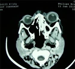
Figure 1: CT Brain
The patient was taken to theatre that same evening for a biopsy and to exclude a fungal infection. She was found to have necrotic, devitalized periorbital soft tissue, with an area of blackening of the left globe medially. Necrotic and devitalized tissue was also found in the mucosa of the ethmoid and maxillary sinuses.
A working diagnosis of mucormycosis was made and a left fronto-ethmoidectomy, left medial maxillectomy and debridement of devitalized tissue was performed. Specimens were submitted to the microbiology laboratory for fungal staining and culture and also to histopathology.
She was admitted to ICU and started empirically on amphotericin B and cefotaxime.
Laboratory Results:
Microbiology
Special fungal stains of tissue samples from the maxillary and ethmoid sinuses, and preseptal area using the potassium hydroxide (KOH) and periodic-acid Schiff (PAS) stains revealed the presence of fungal hyphae in all the specimens. On fungal culture, using both macroscopic (Figure 2a) and microscopic morphology (figure 2b), the mould was identified as a Rhizopus spp.
Figure 2a Macroscopic colonial morphology of fungal isolate
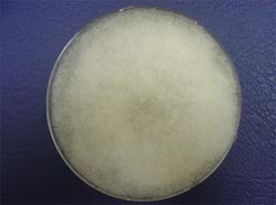
Figure 2b Lactophenol-blue stain demonstrating the microscopic morphology of isolate

Figure 3 shows the results of histopathological examination of the same tissue. The areas of tissue necrosis contained aggregates of aseptate fungal hyphae, which were noted to invade the underlying tissue and vasculature, in keeping with features of mucormycosis. This was further confirmed by special fungal stains (PAS /Grocott’s) which highlighted evidence of invasion by fungal hyphae.
Figure 3 [H&E stain]: numerous fungal elements with tissue invasion and ischaemic necrosis

Question 1: What are the clinical manifestations of mucormycosis?
Answer to Q1
Mucormycosis is an opportunistic fungal infection. The clinical hallmark is tissue necrosis secondary to fungal vascular invasion and thrombosis. Mucormycosis is an aggressive infection, with acute onset, and rapid, relentless progression. Morbidity and mortality, even with combination antifungal and surgical therapy, remains unacceptably high (Skiada et al., 2011).
There are six clinical categories based on the distribution of the anatomical involvement and clinical picture: (1) rhino-orbital-cerebral (craniofacial), (2) pulmonary, (3) gastrointestinal, (4) cutaneous, (5) disseminated and (6) miscellaneous. The various clinical forms are associated with specific underlying host conditions or predisposing factors. Rhino-orbital-cerebral involvement is the most common presentation seen in diabetics with ketoacidosis. Pulmonary disease is typically seen in patients with prolonged neutropaenia or haematopoietic stem cell transplants (HSCT). Malnutrition and prematurity in neonates are factors associated with gastrointestinal presentations. Cutaneous mucormycosis occurs in the setting of burns or penetrating trauma of the skin (Spellberg et al., 2005a).
Rhino-orbital-cerebral Mucormycosis
Infection starts in the paranasal sinuses, and progresses to involve the palate, orbit, face or brain.
Rhino-orbital-cerebral disease occurs mainly in acidotic patients (usually uncontrolled diabetics), but it may also be seen in patients with neutropaenia or organ transplants(Spellberg et al., 2005a).
Rhino-orbital-cerebral mucormycosis has been widely reported as the most common clinical presentation of mucormycosis (Spellberg et al., 2005a, Mantadakis and Samonis, 2009). However, some recent studies have shown the most common site of infection to be pulmonary (Ruping et al., 2010, Skiada et al., 2011). This may be a reflection of the large number of patients with malignancies in the registries of these recent studies.
The clinical picture in the early stages is of a sinusitis. The presence of pyrexia is variable. Initially infected tissue may appear normal. Necrotic eschars in the nasal cavity, on the palate and the face indicate progression of the infection. Erythema, oedema and tissue discolouration precedes the development of the infarcted black tissue. Contiguous spread to the orbit presents as a periorbital cellulitis. Nasal discharge, cavernous sinus thrombosis or decreased level of consciousness may follow with cerebral extension of the infection.
Findings with radiographic imaging are not specific for the diagnosis of mucormycosis, and in early infection may be normal. Hence the importance of surgical exploration and empiric therapy with amphotericin-based antifungals in the high risk patient, while the diagnosis is being confirmed. Findings with imaging include signs of sinusitis, orbital muscle thickening and bony erosions.
Pulmonary Mucormycosis
Pulmonary infection may occur following inhalation, or as a result of lymphatic or haematogenous spread.
Pulmonary mucormycosis is seen most frequently in neutropaenic patients with haematologic malignancies or those who have undergone haematopoetic stem cell transplant (HSCT). Breakthrough mucormycosis infection in patients on azole (including posaconazole) antifungal prophylaxis is increasingly being reported (Mantadakis and Samonis, 2009, Ruping et al., 2010, Skiada et al., 2011)
Clinically and radiographically, pulmonary mucormycosis resembles invasive aspergillosis. It is important to distinguish mucormycosis from aspergillosis infection because voriconazole (the drug of choice for aspergillosis) is not effective against mucormycosis. Persistent pyrexia despite broad-spectrum antibiotics is a common scenario. Other symptoms include dry cough, pleuritic chest pain, dyspnoea and haemoptysis. Angioinvasion results in parenchymal infarction with cavitation and haemoptysis (which can be fatal). Delayed treatment is associated with dissemination of infection (Mantadakis and Samonis, 2009).
High-resolution chest CT scan is the imaging method of choice. Radiographic features include lobar consolidation, nodules and cavitation (Spellberg et al., 2005a)
Gastrointestinal Mucormycosis
Mucormycosis limited to the gastrointestinal tract is rare. It is usually found in malnourished infants and children, and also in patients with haematological malignancies.
Lesions are commonly found in the stomach, colon and ileum and it is seldom diagnosed during life. Symptoms vary and depend on the site affected. These typically include nonspecific abdominal pain and haematemesis. Necrotic ulcers develop, leading to intestinal perforation and peritonitis. Intestinal mucormycosis causes fulminant disease with high mortality.
Cutaneous Mucormycosis
Cutaneous infections account for 16% of all forms of mucormycosis, with an associated lower mortality rate.
Cutaneous mucormycosis is less likely to be associated with severe systemic illness than the other forms. Local predisposing factors include burns, trauma, surgery, and needle sticks. The most common sites involved are the lower and upper extremities, followed by the head and neck, and then the abdomen.
In patients with burn wounds, spread to underlying tissue is common, and usually presents as fever, swelling and changes in the appearance of the wound. In diabetic or immunosuppressed patients, cutaneous lesions may arise at an insulin injection or a catheter insertion site. Mucormycotic gangrenous cellulitis can follow other forms of trauma to the skin. Necrotizing cutaneous mucormycosis has occurred in patients who have had contaminated surgical dressings applied to their skin.
Disseminated Mucormycosis
Disseminated mucormycosis may follow any of the other forms of mucormycosis, but is usually seen in neutropenic patients with pulmonary infection. Less commonly, dissemination can occur from the gastrointestinal tract, or burns, or other cutaneous lesions.
The most common site of spread is to the brain, resulting in abscess formation and infarction. Patients present with sudden onset of focal neurological deficits or coma. CSF microscopic and biochemical investigations are nonspecific and microbiological cultures are sterile. Only imaging techniques have been found to be useful.
Metastatic necrotic lesions have also been found in the spleen, heart, and other organs.
Disseminated mucormycosis has a high mortality rate of up to a 100%.
Question 2: What are the risk factors for development of mucormycosis?
Answer to Q2
Mucormycosis typically occurs in patients with an underlying condition that predisposes them to the development of invasive disease. Cases of mucormycosis in patients without any apparent predisposing conditions are described but these are rare. The predisposing factors typically fall into one of two categories viz. (1) compromised host defences and/ or (2) increased availability of free iron in the serum (Spellberg et al., 2005a).
Compromise of host defences: The classical risk factors such as neutropaenia, use of immunosuppressive agents including corticosteroids, post-transplant all relate to reduced cellular immunity and the inability of phagocytes to effectively clear the organism. Hyperglycaemia and acidosis also impair phagocyte function but the full extent of the predisposition associated with diabetic ketoacidosis has not been fully elucidated. The increased availability of iron in the serum in the ketoacidotic state due to release of iron from binding proteins in the presence of an acidosis likely plays a crucial role in the development of mucormycosis in diabetic patients. Malnutrition impairs host gut immunity; both at an innate and adaptive immune response level, and is thus a risk factor for the development of gastrointestinal mucormycosis. Cutaneous mucormycosis is usually a result of direct trauma and inoculation of the organism. Once again the breach of skin/ mucosal integrity is a compromise in host defences.
Iron is crucial for the growth of most pathogenic organisms and sequestration of iron in the serum is an important host defense mechanism. The use of the iron-chelating agent deferoxamine, contrary to its ability to sequester iron actually predisposes patients to infection with Rhizopus spp. This is because it acts as a siderophore for the organism providing, it with iron from the serum that may previously have been unavailable to the organism (Ibrahim et al., 2008).
Question 3: How is the diagnosis of mucormycosis confirmed in the laboratory?
Answer to Q3
A recent phylogenetic reclassification of fungi means that the order Zygomycetes has been abolished and agents of mucormycosis are no longer synonymous with the term zygomycosis (Hibbett et al., 2007). The six fungal families belonging to the order Mucorales (Mucoraceae, Cunninghamellaceae, Saksenaeaceae, Syncephalastraceae, Thamnidiaceae and Mortierellaceae) are associated with cutaneous and deep fungal infections. The species most frequently identified in cases of mucormycosis are from the Mucoraceae family (Lass-Florl, 2009). In contrast, the Entomophthorales cause infections of the subcutaneous tissues (not angio-invasive disease) in immunocompetent persons in tropical and subtropical regions.
Early diagnosis is essential to allow for optimal management. This requires a high index of clinical suspicion and obtaining suitable samples for laboratory diagnosis. This clinical suspicion must be reported to the clinical laboratory so that the samples are processed appropriately and with urgency in the laboratory. Diagnosis requires the demonstration of fungal invasion of the tissue. In addition species identification is performed by the laboratory.
Lab diagnosis from clinical samples is based on direct microscopy, histopathology, culture and molecular techniques. For rhino-orbital-cerebral disease, scrapings and aspirates from the sinuses, and biopsies of necrotic tissues (nasal, palatal, orbital) must be submitted to the lab. Samples useful for diagnosis of pulmonary mucormycosis include FNA samples, biopsy tissue and bronchoalveolar lavages. Biopsy tissue from patients with gastrointestinal or disseminated infection is required. Cutaneous scrapings/ biopsies are suitable for cutaneous disease. To increase the yield of diagnosis multiple samples must be submitted. Blood cultures are not useful.
Direct microscopic examination
The necrotic material is mounted with 20% potassium hydroxide (KOH). The KOH digests the proteinaceous host cells, making the intact fungal hyphae easier to visualize. Optical brightners such the Calcofluor white stain may be used with or without KOH. Refractile, wide (6-15μm), aseptate hyphae branching at right angles are observed. The hyphae may appear ribbon-like and have focal bulbous dilatation.
These must be distinguished from the hyphae of Aspergillus, which are smaller with parallel walls and dichotomous branching. A positive direct microscopy result from a tissue sample (especially from a sterile site) is clinically significant, even if the culture is negative. Hyphae may be sparse and fragmented in cytological samples (Lass-Florl, 2009).
Histopathology
Fixed tissue is stained with haematoxylin and eosin (H&E) and fungal-specific stains such as Grocott methanamine-silver (GMS) or periodic acid-Schiff (PAS). In addition to the presence of fungal hyphae, angio- and perineural invasion is characteristic. The tissue reaction is usually suppurative but may be granulomatous (Lass-Florl, 2009).
Culture
The Mucorales can be difficult to recover in culture. Homogenising of tissue kills the fungal hyphae and must be avoided. Sabouraud dextrose agar incubated at room temperature and 37 degrees Celsius is recommended. If sample planting is delayed, the sample must not be refrigerated as this reduces organism viability.
Positive culture results from non-sterile sites (for example, sputum) are not diagnostic, as these moulds are ubiquitous and may represent colonisation/ contamination (Lass-Florl, 2009).
Phenotypic identification based on morphologic characteristics can be difficult but is currently widely used in clinical laboratories. The Mucorales are rapid growers that fill the petri plate with fluffy colonies within 2-3 days. Identification is based mainly on the sporangial morphology. In comparison to molecular diagnostic techniques, identification based on morphology can be suboptimal (Dannaoui, 2009).
Serology
Antigen and antibody detection assays require further evaluation and are not currently recommended for clinical use (Lass-Florl, 2009).
Molecular Diagnostics
Molecular diagnostics have a role to play in the identification of Mucorales both directly in tissue samples, and from culture.
Molecular methods (PCR-based and in situ hybridization) have been assessed for the detection of mucormycosis and speciation from unfixed and fixed tissue samples. Various DNA targets, including ribosomal DNA and cytochrome b genes, have been evaluated.
Similarly, the use of various loci and techniques for speciation from cultures, have been reported (Dannaoui, 2009). The Clinical Laboratory Standards Institute provides a guideline for general clinical laboratories on the use of DNA target sequencing for fungal identification (CLSI, 2008). This document focuses on comparative sequence-based identification using the nuclear ribosomal internal transcribed spacer (ITS) region (Balajee et al., 2009).
Molecular techniques have the potential to be more rapid, sensitive and specific than current standard laboratory practices for fungal detection and identification. Ongoing research is required to optimise molecular testing for mucormycosis (Dannaoui, 2009)
Question 4: What is the management of mucormycosis?
Answer to Q4
Principles of management include: (1) Early diagnosis; (2) Early surgical debridement; (3) Appropriate antifungal therapy; (4) Correction of underlying predisposing factor.
Early diagnosis is critical as delayed initiation of therapy has been shown to adversely affect outcome (Chamilos et al., 2008). Diagnosis can be difficult (refer to diagnosis of mucormycosis above) but a high index of suspicion in a patient with an appropriate clinical presentation and an associated risk factor for mucormycosis is critical in making a diagnosis. The pathogenesis of mucormycosis involves angioinvasion with extensive thrombosis and tissue necrosis. The principles of treatment of infectious diseases mandate that source control and adequate penetration of anti-infectives to the site of infection is achieved. Management of mucormycosis thus requires adequate surgical debridement to remove avascular, necrotic tissue and local source control to remove as much of the invasive mycosis as possible. Case series and retrospective analyses have highlighted the role of surgery in reducing mortality although given the devastating disfiguring consequences of aggressive surgery, intraoperative frozen sections to delineate margins of infected tissue has been advocated. The extensive plastic and reconstructive surgery (especially in the rhinocerebral form) required for survivors of mucormycosis cannot be underestimated and is a significant contributor towards further morbidity and mortality.
Antifungal therapy is most often based on the use of a polyene e.g. amphotericin B deoxycholate (AmB). The polyenes remain the cornerstone of antifungal treatment although there are newer agents now available with potential benefit in the treatment of mucormycosis. However the value of newer agents has only experimentally been evaluated as part of combination therapy and are usually added to polyene therapy.
Polyenes: These include AmB and the lipid formulations of AmB which target the ergosterol in the cell membrane creating pores in the membrane. The lipid formulations appear to be a safer option as they can be administered at higher doses for a prolonged period of time with less risk of nephrotoxicity. However comparative efficacy of these formulations has not been directly evaluated in a prospective randomized clinical trial. Liposomal amphotericin B appears to be a better option based on experimental animal models, showing improved survival in mice as compared to AmB (Ibrahim et al., 2003), and a greater than five-fold improved penetration of brain parenchyma as opposed to lipid complex amphotericin B (Groll et al., 2000). Cost considerations are however a limiting factor for the lipid formulations.
Echinocandins: The target enzyme for echinocandins is expressed by Rhizopus oryzae, the commonest cause of mucormycosis, and combination therapy with a polyene has demonstrated benefit in both experimental animal studies (Spellberg et al., 2005b) and in a retrospective study of diabetic patients with rhinocerebral mucormycosis (Reed et al., 2008). Azoles: The only azole with reliable activity against agents of mucormycosis is posaconazole. However low serum levels and inferior efficacy in murine mucormycosis preclude its use as a first-line agent and it should ideally be reserved for salvage therapy or as part of combination therapy (Spellberg et al., 2009).
Adjunctive therapies: These include the use of cytokines (γ-inteferon & granulocyte-macrophage colony-stimulating factor), hyperbaric oxygen and iron chelation therapy. The important role of iron acquisition and metabolism in the pathogenesis of mucormycosis has directed research towards the search for iron chelators other than deferoxamine (see above risk factors). Experimental animal data has demonstrated improved survival when using iron chelators in combination with polyenes (Ibrahim et al., 2008). A randomized, placebo-controlled clinical trial evaluating the safety and efficacy of adjunctive deferasirox has been registered with the Clinical Trials Group (NTC00419770) and results of this study will give further clarity as to the role of iron chelators in the treatment of mucormycosis.
Total duration of antifungal therapy must be individualized for each patient and should be based on clinical response, resolution of radiographic/ clinical findings and correction of underlying predisposition. For patients who require ongoing immunosuppressive therapy it is advisable to continue with secondary antifungal prophylaxis.
Mucormycosis is typically associated with an underlying predisposition (see risk factors above) and thus management of patients must also aim to correct the underlying disease. Aggressive management of diabetic ketoacidosis is imperative to restore homeostasis of both glucose and acid-base status. Management of neutropaenic patients and those on immunosuppressive therapies is more complicated but nevertheless an attempt should be made to correct the neutropaenia, and where possible reduce the dosage of, or stop the immunosuppressive agents.
Acknowledgements: Department of Anatomical Pathology, University of the Witwatersrand for supplying us with the histological specimens and assisting us in taking pictures of the microscopic material.
The patient’s condition deteriorated in ICU and she was found to have developed further proptosis of her left eye. A decision was made to take her back to theatre two days later where the left globe was enucleated, with further debridement of the left orbit, skin, bone and surrounding soft tissue.
Tissue submitted to the microbiology laboratory for fungal staining and culture was still found to have the fungus present. At this point it was decided that no further surgical intervention would be performed and that medical therapy alone would be continued.
She will require extensive reconstructive surgery once her condition improves.
FIDSSA Members can earn CPD points by logging into the secure section of the website and visiting the MyCPD section.
Atlasville, Boksburg
South Africa
2022 © FIDSSA - All rights reserved • Website Terms of Use • Privacy Policy • Powered by E2
Admin login | Website login |
MYMEMBERSHIP®