
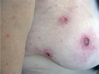
An HIV-negative, 38y old woman staying on a farm near Ficksburg in the eastern Free State presented to the emergency room in Cape Town confused, complaining of a loss of vision and a rash for 1 week. She had been seen by a local practitioner 2 days previously, who had started her on oral doxycycline and referred her to the eye clinic. No other history was available. Ophthalmology review found normal vision and no evidence of retinopathy or uveitis. She had a low grade fever, multiple lesions on the breast (figure 1) and 2 similar lesions on her back, with a maculopapular rash on her trunk, abdomen and limbs, involving the palms but not soles. There was no lymphadenopathy or organomegaly, she was disorientated in time, place and person, but had no cranial or peripheral nerve lesions.
Her platelet count was 140 x 109/L, but the rest of her full blood count, liver function and renal function tests were normal. Lumbar puncture revealed:
During the consultation, an anguished telephone call was received from relatives on the farm in Ficksburg asking if her illness could be related to the outbreak of Rift Valley Fever in farms around where she was staying.
Question 1: What is the most likely diagnosis, what investigations would you send to confirm the diagnosis and how would you treat it?
Answer to Q1
Spotted Fever Group (SFG) Rickettsiosis, most likely Mediterranean Spotted (Boutonneuse) Fever subtype caused by Rickettsia conorii infection
Rickettsioses are a group of zoonotic infections caused by intracellular gram negative bacteria. Rickettsial diseases are subdivided into:
SFG rickettsioses are the commonest rickettsial infections in Africa. ATBF, by far the commonest rickettsial infection seen in returning travellers from Africa is caused by infection with Rickettsia africae, transmitted by the Amblyomma tick, a game and cattle tick found on the vegetation in the bush. Visitors to game reserves, hunters and ecotourists traveling during late summer months appear to be at heightened risk, although a recent study concentrating to South Africa has shown that travellers are at heightened risk of contracting ATBF during the winter months (June-Sept). MSF in contrast is caused by R. conorii, transmitted by the brown dog tick and is less commonly associated with travel.
ATBF and MSF are characterised by an acute febrile illness with the organisms targeting vascular endothelial cells inducing a vasculitis. After an incubation period of 5-7 days, the classic triad of eschar (as demonstrated in figure 1), fever and rash occur. Headache and myalgia are common accompanying symptoms. Distinguishing features of ATBF include an association with multiple inoculation eschars, regional adenopathy and the rash may be vesicular in nature, rather than maculopapular rash. In contrast, eschars in MSF are usually single and often hard to find in remote sites such as the hairline. This case is unusual in having multiple eschars. MSF may also be characterised by more serious illness. A study of 199 cases of serologically-proven MSF documented a severe form of the disease in 6% of patients. In the same study, meningism was noted in 11% of patients and altered mental state in 10%. There are multiple case reports documenting various combinations of meningoencephalitis, with CSF characteristics similar to our case, i.e. white cell pleocytosis, mildly raised protein and normal CSF glucose.
Rickettsioses are usually associated with lymphopenia, thrombocytopenia, raised CRP and moderately elevated liver enzymes. Diagnosis should rely primarily on the clinical findings of eschar plus the febrile illness, and this should prompt treatment. Laboratory diagnosis is secondary as frequently even specific rickettsial serology may only become positive after 5-7 days of the illness or by demonstration of an increase in specific titres on acute and convalescent sera using IgM. PCR of blood or eschar biopsy is able to differentiate between R conorii and R africae. The Weil-Felix agglutination test is neither specific nor sensitive and should no longer be used.
Oral doxycycline 100mg bd for 7-10 days is the drug of choice, although if unable to take oral therapy as iv doxycycline is unavailable, ivi ciprofloxacin or if not available ivi chloramphenicol are alternatives. Clinical response to macrolides may be suboptimal. Complications to MSF do occur especially in persons in whom treatment is delayed. In contrast to MSF, fatalities from ATBF are very rare.
Question 2: What is the main differential diagnosis in this case?
Answer to Q2
With the risk exposure of living on a farm in the Free State, a rash and confusion, the main differential diagnosis to rule out in this case would be Crimean Congo Haemorrhagic Fever (CCHF). Entertaining a diagnosis of CCHF has major implications in terms of likelyhood of potential severity of disease and in terms of infection prevention and control practices to isolate the patient once the diagnosis is suspected.
The incubation period of CCHF virus (1-3 days following tick exposure) is shorter than that of the rickettsiae, although as a proper history is lacking in this case, it would not help to differentiate the two. Generally, the bite of Hyalomma ticks which carry CCHF do not cause eschars. The pre-haemorrhagic period, characterised by sudden onset of fever, rigors, meningism and gastrointestinal symptoms lasts 3-6 days, which may be followed by the haemorrhagic period. Abnormal liver function tests are almost invariable in CCHF by the time the haemorrhagic period is reached, as is thrombocytopaenia. The normal LFTs and platelet count just under the normal range in our patient make the diagnosis of CCHF unlikely.
Question 3: Why is this illness not characteristic of Rift Valley Fever?
Answer to Q3
Rift Valley Fever is predominantly a disease of livestock, transmitted to humans through direct or indirect contact with blood or tissues of infected animals. Farmers, abattoir workers and veterinarians are at increased risk. Transmission may also occur via mosquito bites or the ingestion of unpasteurized milk. Ticks are not vectors for RVF and human-to-human transmission has not been documented.
After an incubation period of 2-6 days, there may be a sudden onset of flu-like illness with our without myalgia and symptoms remain for 4-7 days. Eschars are not a feature of RVF. Rarely, the illness may be complicated by severe manifestations, haemorrhagic fever and hepatitis which occur in < 1% of cases, has a case fatality of ~50%. meningoencephalitis (< 1%) and ocular involvement (~0.5%) are also rare. Ocular involvement commonly begins 1-3 weeks after the start of the illness and manifests as blurred or decreased vision or field defects secondary to macular or peri-macular retinitis. Up to half of all cases may lead to permanent damage.
A clinical diagnosis of Mediterranean Spotted Fever was made and rickettsial serology was sent. The patient was switched to intravenous ciprofloxacin and she was sedated overnight to control aggressive behaviour. By day 2, she was no longer confused and was able to give a history of multiple ticks having been seen on her body after cuddling the farm’s dog 6 days prior to the onset of the rash. She had had no contact with other farm animals. She admitted to having been nauseated and vomiting for 2 days prior to admission, unable to keep her oral doxycycline down. She was discharged on day 3, back on oral doxycycline and made an uneventful recovery. Acute and convalescent serology confirmed rickettsial infection.
FIDSSA Members can earn CPD points by logging into the secure section of the website and visiting the MyCPD section.
Atlasville, Boksburg
South Africa
2022 © FIDSSA - All rights reserved • Website Terms of Use • Privacy Policy • Powered by E2
Admin login | Website login |
MYMEMBERSHIP®