ABOUT FIDSSA

Case of the Month

March 2017

Frans Radebe, NICD/NHLS & Dr Marcille Le Roux, University of Limpopo (Medunsa)

A 29-year old man presented to a dedicated STI clinic with discrete, nontender skin lesions on the pubis/groin consisting of multiple rounded, flesh-colored dome-shaped, pink waxy painless papules approximately 5 mm in diameter widely disseminated without plaque formation. The papules were umbilicated and contained a plug. There were no lesions elswewhere. The patient complained of irritation due to inflammation and stress cause by the infection which he had been experiencing for the last two months. The patient was HIV-infected with a CD4 cell count of 200 cells/mm3. A clinical diagnosis of molluscum contagiosum was made and the patient managed as per the national STI treatment guidelines, 2014.

Figure: A patient discrete dome-shaped lesions manifestations of Molluscum contagiosum

Question 1 - Describe the aetiology, epidemiology and clinical features of Molluscum contagiosum

Answers to Q1

Molluscum contagiosum (MC) is caused by a DNA poxvirus of the same name, and is largely a disease of humans. Infection follows contact with infected persons or contaminated objects. MC virus transmission through direct skin contact between children sharing a bath and between athletes sharing gymnasium equipments and benches has been documented. Between 5-18% of children show signs of clinical disease, increased in patients with HIV infection, with severity inversely proportional to CD4 count. In the general population the prevalence of MC is 33% in developed countries. Even though reinfection is common, the virus is not strongly immunogenic and infrequently induces antibody formation that can be detected by several serological tests such as complement fixation test. It was first described by Bateman in 1817, its viral nature in 1905 by Juliusburg and its infectious nature by Paterson in 1941. Four types of MC have been identified by restrictive endonuclease analysis of the viral genome namely MC I, II, III and IV.

Type I causes 96.6% of infections in non-HIV-infected individuals, while type II causes 60% of infections in HIV-infected patients. Types III and IV are rare. It is more prevalent in tropical areas and transmission may be related to poor hygiene, warmth and humidity. It is more common in whites than other races and more common in males than females. It peaks among pediatric age group due to casual contact and among young adults during sex. Use of school swimming pools correlates with childhood infections. MC infection normally resolves without therapy within 6-9 months, but may persist for 3-4 years.

Infection in adults is usually sexually transmitted with lesions limited to the perineum, lower abdomen or buttocks. Complications of MC are mainly irritation, inflammation and secondary infection. Lesions on eyelids are associated with follicular or papillary conjunctivitis. Bacterial super-infection in HIV-infected patients is associated with Staphylococcus aureus and Pseudomonas aeruginosa with abscess formation. MC may be widespread, persistent and atypical in immunocompromised patients.

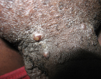
Patients on immunosuppressive medication may have more extensive infections, which are difficult to mangage. Patient education as to the benign nature of the disease is important to allay the distress it can cause in patients or parents of infected children. It is unclear whether condoms and other barrier methods provide adequate protection against transmission, thus safe sex and abstinence in adolescents and adults should be encouraged. MC can occur on penis, scrotum, inner thigh or any other parts of the body.

Question 2: What is the diagnosis and other investigations would you do to confirm the diagnosis?
Answer to Q2: Differential diagnosis

Early lesion may be confused with genital warts or herpes but unlike herpes they are painless. MC can be mimicked and confused with other infections in the immunocompromised, such as cutaneous cryptococcosis, histoplasmosis, aspergillosis and cutaneous coccidioidomycosis. Basal cell carcinoma, condyloma accuminatum, pearly penile papules, varicella-zoster virus and keratosis pilaris also need excluding. The distinctive clinical feature of MC is central umbilication of the dome-shaped lesion. Histologic or microscopic confirmation of MC may be necessary simply done by expressing the pasty core of the lesion by crushing the lesion between two microscope slides to release particulate MC virions. This can then be stained by Giemsa, Grams, crystal violet, Papanicolaou test, biopsy of the lesion reveal intracytoplasmic inclusion bodies typical of molluscum bodies or Henderson-Paterson bodies. Hematoxylin & Eosin stained histologic section also reveals cup-shaped indentation of the epidermis into the dermis with purple to red stained Henderson-Paterson bodies shown by the presence of intracytoplasmic, eosinophilic, granular inclusions within the keratinocytes of the basal and granular layers of the epidermis. Also diagnostic are lobules containing hyalinized molluscum bodies.

Question 3: How would you treat this patient?
Answer to Q3

In healthy patients who are not immunocompromised, MC infection is self-limited and heals spontaneously after several months. The most important precautionary measure in both children and adults is to reduce autoinoculation or transmission to close contacts and to improve the clinical appearance. Topical application of medications such as Podophylin, Potassium hydroxide, Tretinoin cream and Cantharidin, radiation therapy and/or surgery is recommended. These include cryotherapy or curettage of individual lesions in adults. Therapy with lasers, imiquimod and antiviral therapy or combinations of both have been successfully used. Treatment may result in scarring or post-inflammatory pigment changes. Multiple treatment sessions may be necessary because of the recurrence of treated lesions and/or appearance of new lesions due to autoinoculation. Once the lesions have completely resolved MC virus doesn’t remain latent. I can be completely cured. However, there is no permanent immunity to the virus one may get infected again upon contact to an infected person.
Outcome of the case

The patient was educated about the diagnosis and the benign nature of the disease, the risk of autoinoculation or infection of others and the therapeutic options available at the clinic and elsewhere. Good handwashing and limiting the physical contact with infected areas of skin to reduce transmission was emphasized including safe sex and abstinence since concomitant infection with other STIs may be a possibility. He was encouraged to return to the clinic for follow-up treatment and to monitor recurrent infections if any.

Further reading:
  1. Scholz J, Rosen-Wolff A, Bugert J, et al. Epidemiology of molluscum contagiosum using genetic analysis of the viral DNA. J Med Virol. Feb 1989; 27 (2): 87-90.
  2. Choong KY, Roberts LJ. Molluscum contagiosum, swimming and bathing: a clinical analysis. A J Dermatol. May 1999; 40 (2): 89-92.
  3. Laxmisha C, Thappa DM, Jaisankar TJ. Clinical profile of molluscum contagiusum in children vs adults. Dermatol. Online J Dec 2003 ; 9 (5): 1
  4. National guideline for the management of molluscum contagiosum. Sex Transm Aug 1999; 75 Suppl 1: S80-1
  5. Buller RM, Burnett J, Chen W, Kreider J. Replication of molluscum contagiosum virus. Virology. Nov 10 1995; 213 (2): 655-9
  6. Cribier B, Scrivener Y, Grosshans E. Molluscum contagiosum: histologic patterns and associated lesions. A study of 578 cases. AM J Dermatopathol. Apr 2001; 23 (2): 99-103

Earn CPD Points

CPD QUESTIONS

FIDSSA Members can earn CPD points by logging into the secure section of the website and visiting the MyCPD section.